Figure 1. RPGR interacts with NPHP4. A:
Bovine
retinal lysate was subjected to IP using RPGR (A)
antibody or control IgG (IgG from pre-immune bleed of rabbits) followed
by immunoblotting using anti-NPHP4 antibody. The input lane represents
20% of the protein used for IP. B: The NPHP4-RPGR complex is
disrupted in Rpgr-ko retinas: Protein lysates from wt
or Rpgr-ko retinas were immunoprecipitated with RPGR antibody
and analyzed by immunoblotting with NPHP4 antibody. Lanes are
indicated. C: Schematic representation of the primary structure
of NPHP4. BD represents the binding domain. D: Interaction of
GST-RPGR with 35S in-vitro translated NPHP4 was analyzed by
GST pull-down assay, as described in the experimental procedures.
Purified GST moiety was used as control. The lower panel shows
Coomassie blue stained gel of the GST-RPGR and GST protein (asterisks)
used in the assay. E: The GST pull-down assay was performed
using GST-RPGR and 35S-labeled deletion mutants of NPHP4.
The lower panel shows Coomassie blue staining of the GST-RPGR protein
used in this assay. Molecular markers in kDa are shown on the left.
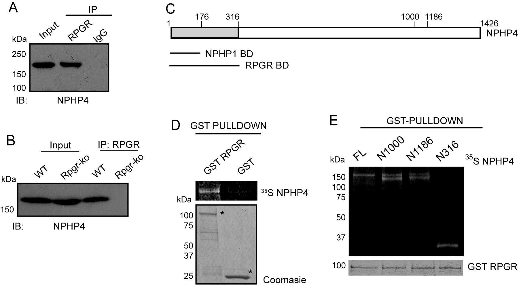
 Figure 1 of Murga-Zamalloa, Mol Vis 2010; 16:1373-1381.