Figure 7. Drawing of a Müller cell with
its nucleus and vimentin cytoskeleton showing the proposed hypothesis
of how these cells may undergo nuclear division following retinal
injury. Step 1, after retinal detachment the nucleus migrates to the
outer nuclear layer (ONL); Step 2, vimentin filaments accumulate around
the nucleus after which the nucleus undergoes mitosis; Step 3, one
nucleus migrates back to the inner nuclear layer (INL); Step 4, the
nucleus remaining in the ONL either moves to the subretinal space and
contributes to the formation of a glial scar (Step 5) or remains in the
retina as a de-differentiated cell (Step 6).
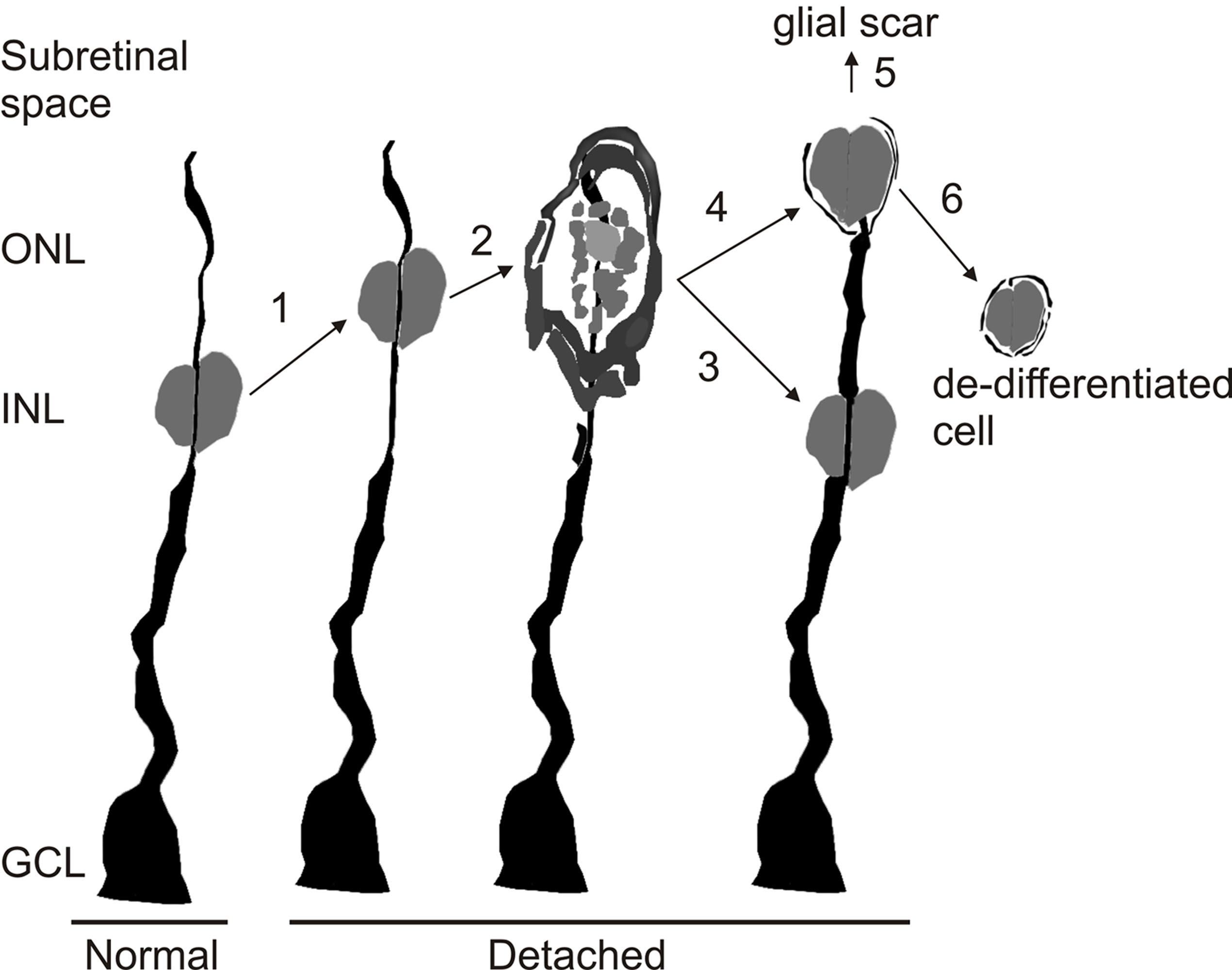
 Figure 7 of Lewis, Mol Vis 2010; 16:1361-1372.