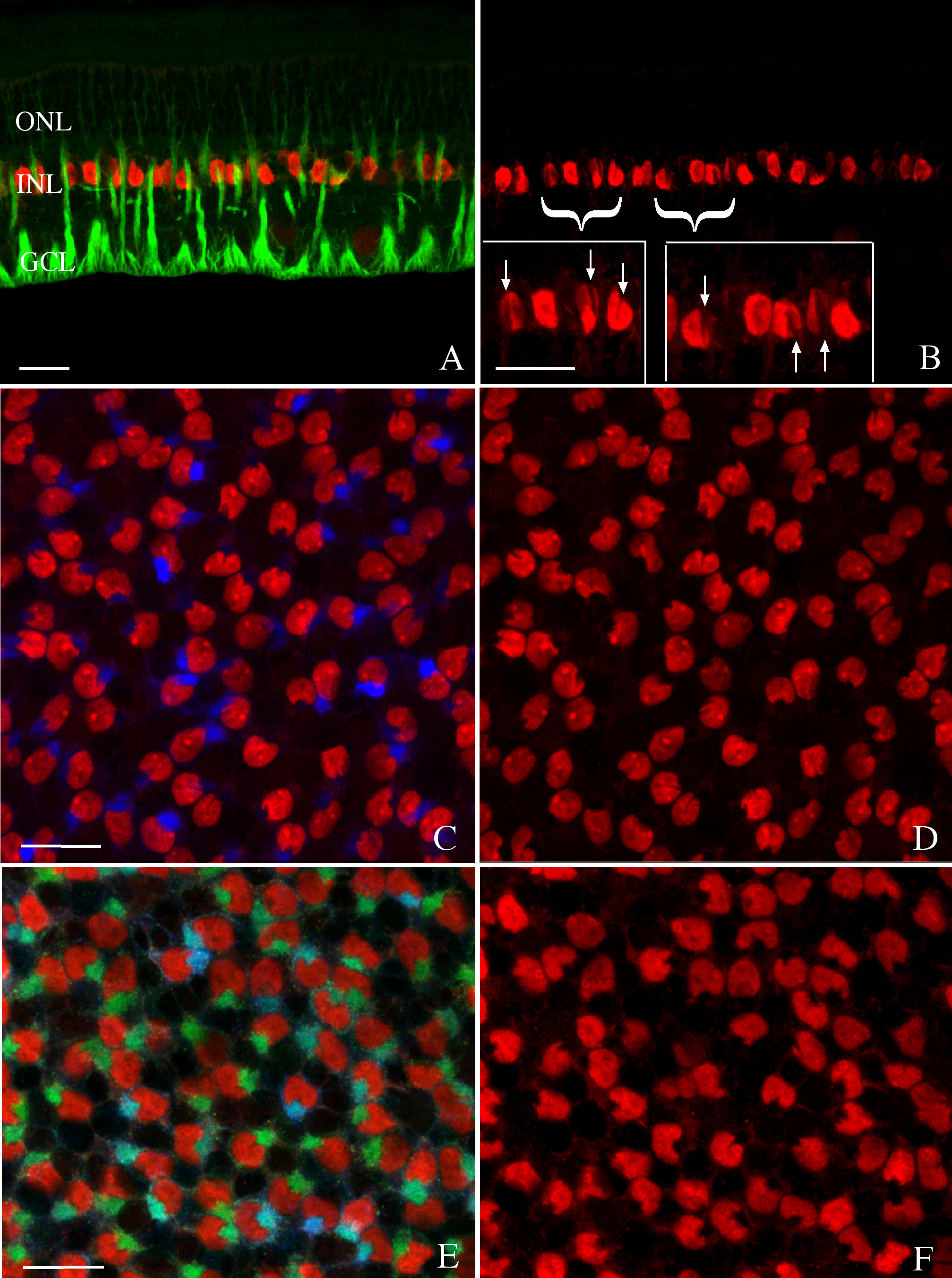
Figure 6. Laser scanning confocal images
of radial sections (A, B) and wholemounts (C- F)
of
control rabbit retina labeled with anti-bromodeoxyuridine (BrdU;
red), anti-vimentin (green), and anti-glial fibrillary acidic protein
(GFAP; blue) from eyes injected with 5-fluorouracil (5-FU). The
anti-BrdU detects the 5-FU, which is present in all Müller cell nuclei
and illustrates that the notch is visible in most of them (A-F).
In
addition, GFAP filaments are present in only some of the notches (C,
D), while vimentin filaments appear to be present in all of the
notches (E, F). The images in the right panels are the
same as those on the left but illustrate only the BrdU labeling. The
insets in B are higher magnifications of the regions marked
with the brackets to more easily visualize the notch (arrows).
Abbreviations: GCL represents ganglion cell layer; INL represents inner
nuclear layer; ONL represents outer nuclear layer. The scale bars are
equal to 20 µm.

 Figure 6 of Lewis, Mol Vis 2010; 16:1361-1372.
  Figure 6 of Lewis, Mol Vis 2010; 16:1361-1372.