Figure 5. Three images illustrating the
indentation or “notch” within a single Muller cell nucleus. A, B:
Laser
scanning confocal images of rabbit retina detached for 3 days and
labeled with both anti-bromodeoxyuridine (BrdU; red) and anti-vimentin
(green). B is the same image as shown in A, with the
two colors separated to better visualize the vimentin filaments (green)
and the notch in the (single) nucleus (red, inset). C, D:
Electron
micrographs of normal rabbit (C) and 50-day detached
cat (D) retinas showing a notch within Muller cell nuclei
(arrows). Note the Müller cell nucleus in D now resides among
the dark photoreceptor nuclei after detachment. Abbreviations: ONL
represents outer nuclear layer; INL represents inner nuclear layer; GCL
represents ganglion cell layer. Scale bars equal to 10 µm A, B;
5
µm C, D.
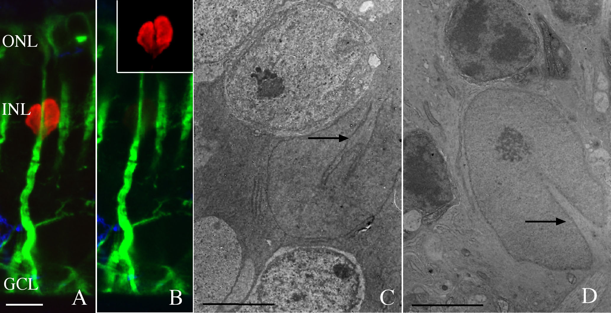
 Figure 5 of Lewis, Mol Vis 2010; 16:1361-1372.