Figure 4. Laser scanning confocal images
of rabbit retinas detached for 4 days and labeled with
anti-bromodeoxyuridine (BrdU; red), anti-phosphohistone H3 (green), and
anti-vimentin (blue). BrdU was injected intravitreally on day 3.
Anti-phosphohistone H3 labeling of mitotic cells is only observed in
the outer nuclear layer (ONL). Some, but not all, of the
anti-phosphohistone H3-labeled cells are also labeled with anti-BrdU (A,
B). The insets in A and B are cropped from the
same image but without the red BrdU channel to more easily visualize
the phosphohistone H3 labeling (long arrows point to the same nucleus
without the BrdU labeling). Many of the anti-phosphohistone H3-labeled
nuclei are surrounded by an accumulation of vimentin filaments (A,
C, D, short arrows). Figures C and D are
the same image but digitally rotated by tilting the z-stack in
different directions to reveal the three-dimensional architecture of
the vimentin filaments around the mitotic cell. The top and bottom
images of the z-stack were omitted to visualize the anti-phosphohistone
H3 labeling within this region. Abbreviations: INL represents inner
nuclear layer; IPL represents inner plexiform layer; GCL represents
ganglion cell layer, OLM represents the outer limiting membrane, ONL
represents the outer nuclear layer. Scale bars are equal to 50 µm (A,
B) or 20 µm (C, D).
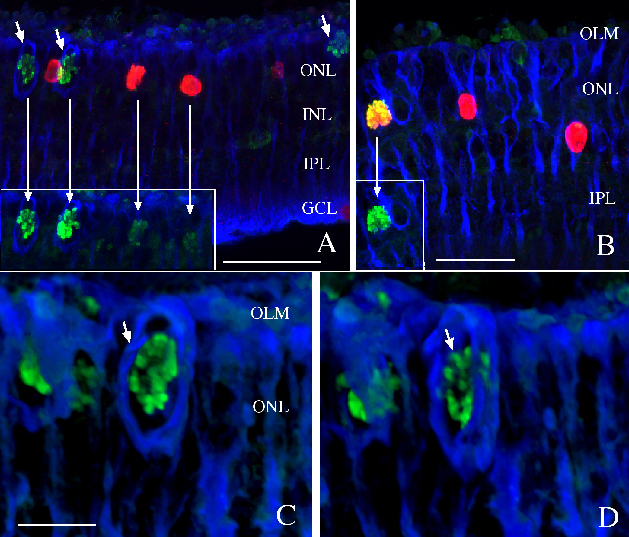
 Figure 4 of Lewis, Mol Vis 2010; 16:1361-1372.