Figure 3. Laser scanning confocal images
of rabbit retinas detached for 3 weeks and labeled with
anti-bromodeoxyuridine (BrdU; red), anti-vimentin (green), and
anti-S100 (blue). BrdU was injected intravitreally on day 3. Many
anti-BrdU-labeled nuclei are present throughout the retina, some of
which are co-labeled with anti-S100, giving them a purple appearance,
while many others do not label with anti-S100 and appear red (arrows).
The areas labeled with all three antibodies appear white (A,
arrowhead). The images in B and D are the same images
as A and C without the red (anti-BrdU) channel.
Abbreviations: GCL represents ganglion cell layer; INL represents inner
nuclear layer; ONL represents outer nuclear layer. The scale bars are
equal to 50 µm.
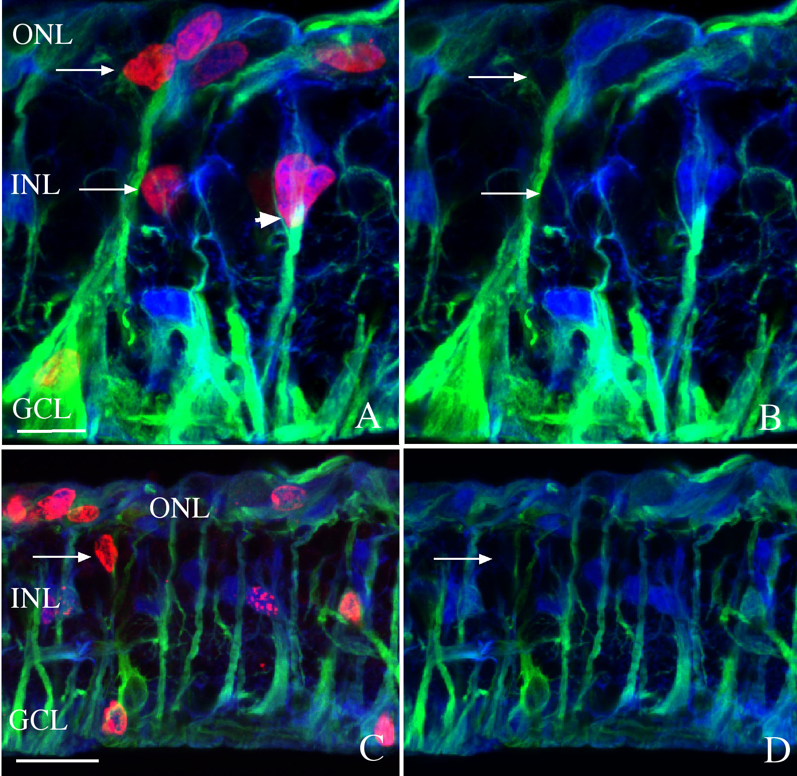
 Figure 3 of Lewis, Mol Vis 2010; 16:1361-1372.