Figure 2. Laser scanning confocal images of rabbit retinas detached for 4 days labeled with anti-bromodeoxyuridine (BrdU; red) and anti-vimentin
(green). The z-stack of images is projected in three dimensions at varying contrasts, illustrating that the BrdU-labeled nuclei
appear to be associated with only one Müller cell process. A shows two nuclei lined up directly with a single Müller cell “stalk” of intermediate filaments (arrows). The image in B is rotated slightly so that the lower nucleus in the INL appears off to the side of the major intermediate filament bundle.
Abbreviations: GCL represents ganglion cell layer; IPL represents inner plexiform layer; INL represents inner nuclear layer;
ONL represents outer nuclear layer; OLM represents outer limiting membrane. The scale bar is equal to 20 µm.
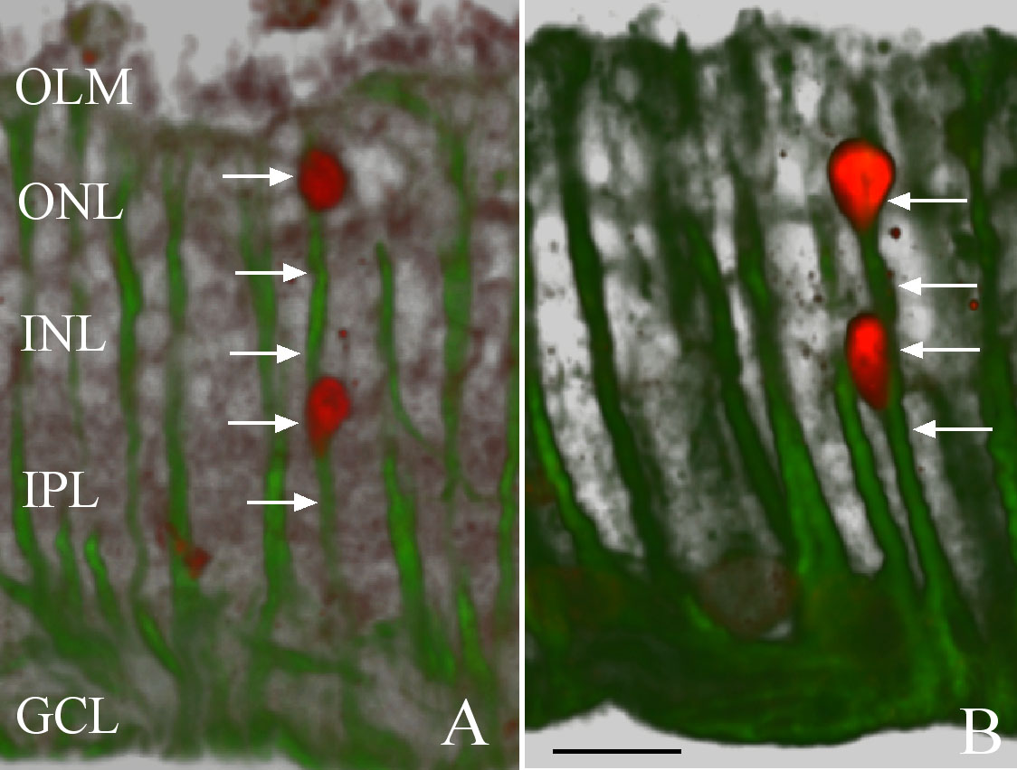
 Figure 2 of
Lewis, Mol Vis 2010; 16:1361-1372.
Figure 2 of
Lewis, Mol Vis 2010; 16:1361-1372.