Figure 1. Laser scanning confocal images
of control (A) and detached (B-F) rabbit retinas
labeled with anti- bromodeoxyuridine (BrdU; red), anti-vimentin
(green), and isolectin B4 (blue). BrdU was injected intravitreally on
day 3. In control retina (A; 4 h after BrdU injection into the
normal eye), no BrdU is detected and anti-vimentin labeling of Müller
cells extends from their endfeet into the outer nuclear layer (ONL). At
3 days after detachment (4 h after intravitreal injection of BrdU),
anti-BrdU labeling is present in many Müller cell nuclei in the inner
nuclear layer (INL), and anti-vimentin labeling in Müller cells spans
the entire width of the retina. At 4 days after detachment (24 h after
BrdU injection), BrdU-labeled nuclei frequently appear in radial
columns across the retina (C, arrows). In some cases, two nuclei
can be observed directly adjacent to one another (D, arrows).
Anti-BrdU-labeled nuclei are also observed directly adjacent to
anti-vimentin-labeled Müller cell processes extending into the
subretinal space (E, brackets). At 7 days after detachment, many
anti-BrdU-labeled Müller cells are observed in large subretinal glial
scars that are also labeled with anti-vimentin (F, brackets).
The isolectin B4 labels the stellate processes of the microglia in the
inner plexiform layer (IPL) in the control retina (A), but after
detachment these cells round-up and migrate throughout the retina and
into subretinal glial scars (B-F). The isolectin B4 also
labels macrophages in the subretinal space (B-F). Some
macrophages (in the subretinal space) and microglia (in the retina) are
labeled with anti-BrdU (D, E, F, arrowheads).
Abbreviations: GCL represents ganglion cell layer; OLM represents outer
limiting membrane. Scale bars are equal to 50 µm.
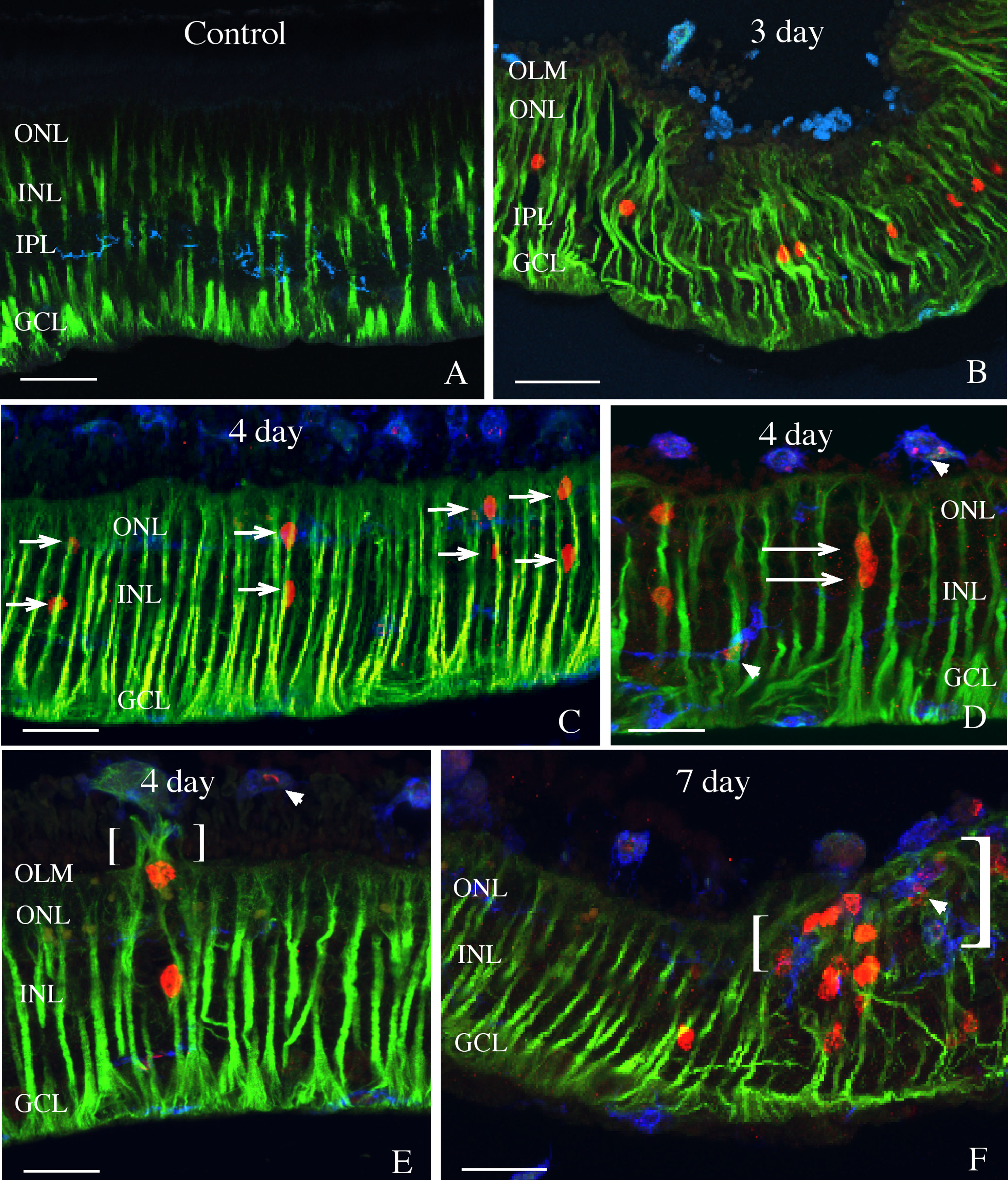
 Figure 1 of Lewis, Mol Vis 2010; 16:1361-1372.