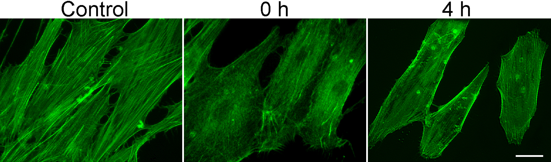
Figure 3. Actin staining in human
trabecular meshwork cells. Cells were treated with 1 mM H
2O
2
for 30 min and were fixed immediately after (0 h) or 4 h later (4 h),
and stained with Alexa Fluor 488-phalloidin. Cells untreated were used
as controls. Results showed a significant reduction in actin stress
fibers upon H
2O
2 treatment, a previously
documented cell response [
22]
to
oxidative stress. Scale bar represents 20 µm.
 Figure 3 of Shyam, Mol Vis 2010; 16:122-129.