Figure 7. Increased optic atrophy type 1 (OPA1) expression blocks mitochondrial fission in differentiated retinal ganglion cell (RGC)-5
cells exposed to elevated hydrostatic pressure (30 mmHg) for three days. Mitochondria are stained with MitoTracker Red. A, C: Non-pressurized control cells without transfection. B, D: Non-pressurized control cells transfected with AAV2 Null. E,G: Pressurized cells transfected with AAV2 Null. F,H: Pressurized cells transfected with AAV2-WT mOPA1. High magnification showed that increased OPA1 expression blocked mitochondrial
fission in pressurized cells that are counterstained with Hoechst 33342 (blue). Scale bar indicates 10 µm (A,C, F, H).
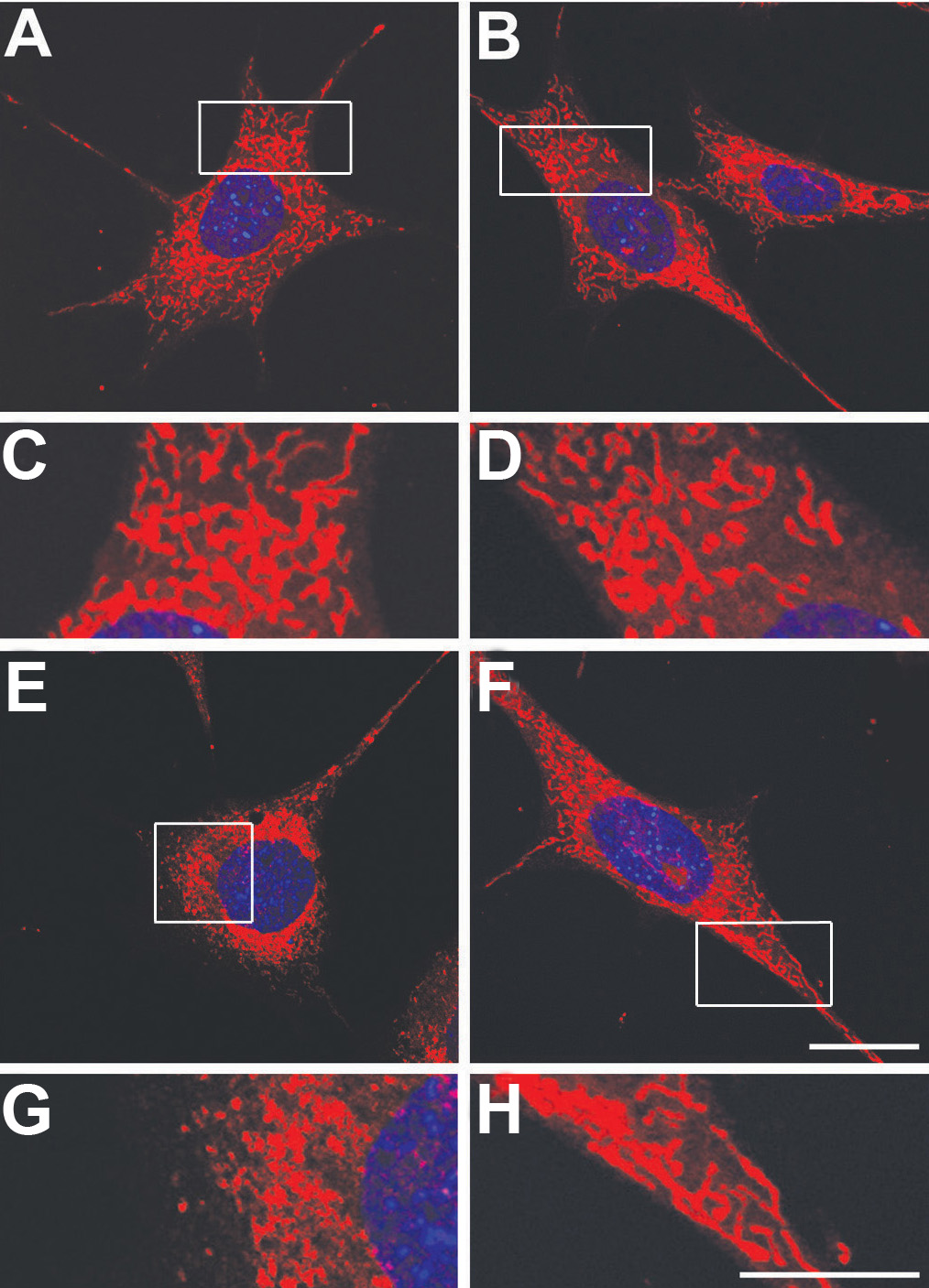
 Figure 7 of
Ju, Mol Vis 2010; 16:1331-1342.
Figure 7 of
Ju, Mol Vis 2010; 16:1331-1342.