Figure 4. Retinal ganglion cell (RGC) survival in nine-month-old glaucomatous DBA/2J mice following transfection with adeno-associated
virus serotype 2-cytomegalovirus-green fluorescent protein (AAV2-CMV-GFP) or adeno-associated virus serotype 2-wild type (AAV2-WT)
optic atrophy type 1 (mOPA1). The representative flatmount photomicrographs are from the middle retina following retrograde
labeling with FluoroGold. A: Non-glaucomatous C57BL/6 mice transfected with AAV2-CMV-GFP; n=4 retinal flatmounts/mice/group. B: Glaucomatous DBA/2J mice transfected with AAV2-CMV-GFP; n=4 retinal flatmounts/mice/group. C: Glaucomatous DBA/2J mice transfected with AAV2-WT mOPA1; n=7 retinal flatmounts/mice/group. D: The quantitative analysis of RGC survival. Data represent the mean±SD *Significant at p<0.05 compared with non-glaucomatous
C57BL/6 mice transfected with AAV2-CMV-GFP or **Significant at p<0.05 compared with glaucomatous DBA/2J mice transfected with
AAV2-CMV-GFP. Scale bar indicates 50 µm (C-E).
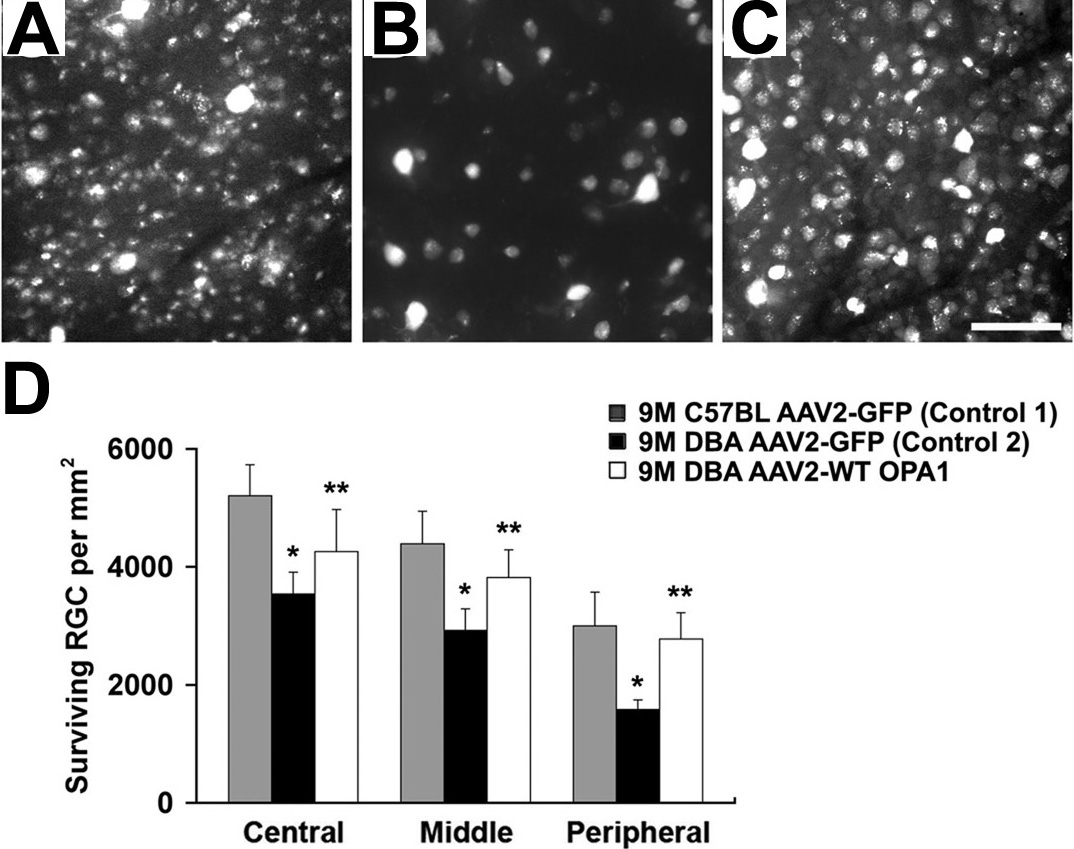
 Figure 4 of
Ju, Mol Vis 2010; 16:1331-1342.
Figure 4 of
Ju, Mol Vis 2010; 16:1331-1342.