Figure 2. Adeno-associated virus serotype 2-cytomegalovirus-green fluorescent protein (AAV2-CMV-GFP) labeling in the retina and optic
nerve of six-month-old DBA/2J mice. A: Normal appearance of retinal cross section. B: The ganglion cell layer (GCL) from the retinal flatmount. C: Optic nerve fiber staining in a whole eye section after triple labeling with AAV2-CMV-GFP (green), neurofilament (red),
and glial fibrillary acidic protein (GFAP; blue). D: The entire dendritic tree (arrowheads) and cell body (arrow) of retinal ganglion cell (RGC) labeled with AAV2-CMV-GFP (green)
and DiI (red). E: Enlargement of the retinal section shown in panel C to illustrate staining in the RGC somas (arrows) and axons (arrowheads) in the GCL. F: Enlargement of the optic nerve head shown in panel C demonstrates triple labeling with AAV2-CMV-GFP (green), neurofilament (red), and GFAP (blue). Abbreviations: IPL represents
inner plexiform layer; GCL represents ganglion cell layer; CA represents central artery. Scale bar indicates 20 µm (A, D-F), 50 µm (B), and 100 µm (C).
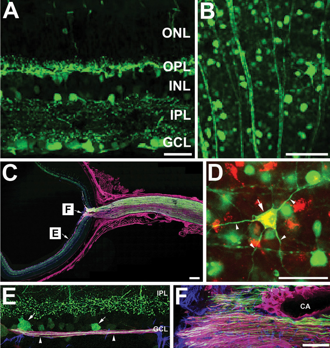
 Figure 2 of
Ju, Mol Vis 2010; 16:1331-1342.
Figure 2 of
Ju, Mol Vis 2010; 16:1331-1342.