Figure 9. Confocal laser corneal
microscopy images of the rats' corneal stroma ten weeks after
transplantation. A: Normal corneal stromal cells were well
arranged. B: Neovascularization (arrow) can be seen in the
anterior matrix with the proliferation of stromal cells in the control
group. C: Stromal cells proliferated and inflammatory cells
existed in the anterior stroma of the AM group. D: Stromal
cells also proliferated while a few neovascularizations appeared in the
stroma of the induced MSC group.
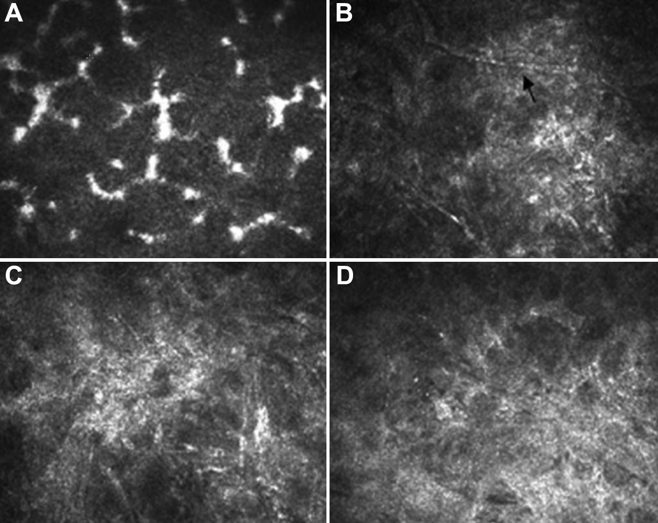
 Figure 9 of Jiang, Mol Vis 2010; 16:1304-1316.