Figure 8. Confocal laser corneal
microscopy images of the rats' corneal epithelium ten weeks after
transplantation. A: Epithelium cells were well arranged and
integrated closely in the normal cornea. B: The structure of
epithelial cells was not clear with ambiguous boundary in the control
group. C: The epithelial cells of the AM group were disarranged
with an enhanced reflection and cavities (arrow). D: The
epithelial cells of the induced MSC group were integrated with clear
boundaries.
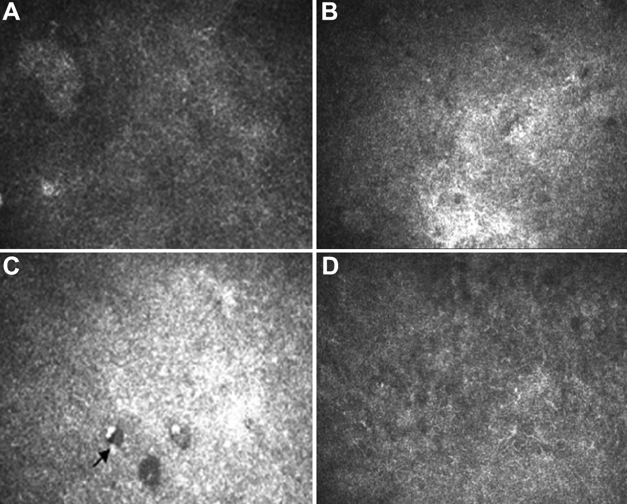
 Figure 8 of Jiang, Mol Vis 2010; 16:1304-1316.