Figure 6. H&E staining of four groups
ten weeks after transplantation. A: Control group: Epithelial
structure was disordered, and numerous inflammatory cells and
neovascularization emerged. B: AM group: Epithelium was
integrated and some inflammatory cells infiltrated in the stroma. C:
Non-induced
MSC group: Incomplete epithelial hyperkeratosis and
inflammatory cell infiltration. D: Induced MSC group:
Epithelium was intact with a few lymphocytes and neovascularization
infiltration. The magnification was 200×.
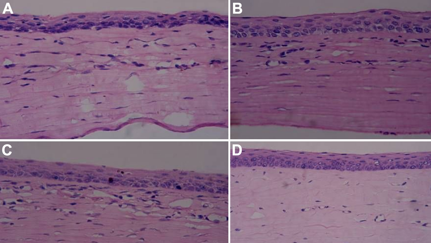
 Figure 6 of Jiang, Mol Vis 2010; 16:1304-1316.