Figure 4. Surgical effects of different
treatments in the rat corneal alkali burn model. A, B:
Control group (A). Four weeks after transplantation, corneal
epithelial defect size > 1/2 quadrant and neovascularization grew
rapidly and involved 1/2 quadrants, with ulcer emerged (arrow) (B).
The
new blood vessels grew into the whole cornea with extremely severe
opacity ten weeks after transplantation. C, D: AM group
(C). Moderate to severe corneal opacity four weeks after
transplantation (D). Corneal neovascularization grew into nearly
3/4 quadrants ten weeks after transplantation. E, F:
Non-induced MSC group (E). Moderate corneal opacity with local
stromal scar was seen four weeks after surgery (F).
Conjunctivalization with apparent neovascularization was found in
1/2–2/3 quadrant ten weeks after surgery. G, H: Induced
MSC group (G). The corneal opacity was mild and there were no
signs of conjunctival epithelium growing into the cornea four weeks
after transplantation (H). Cornea kept transparent and the
neovascularization was within 2 mm from limbal ten weeks after surgery.
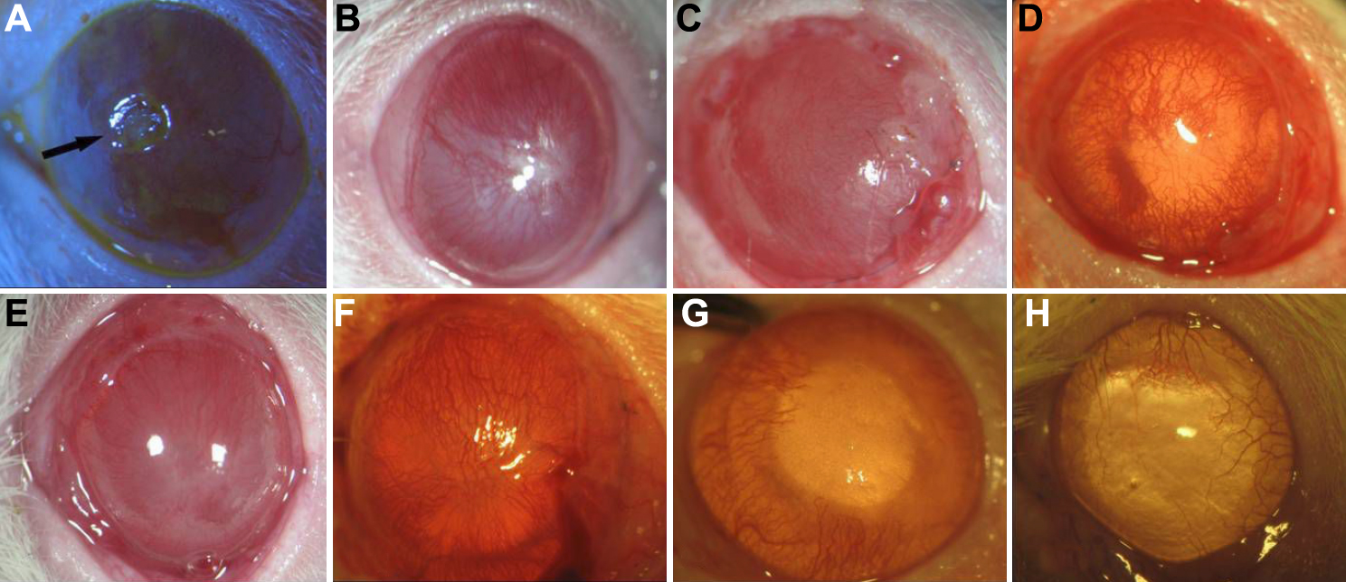
 Figure 4 of Jiang, Mol Vis 2010; 16:1304-1316.