Figure 3. Induced MSCs on the amniotic
membrane (AM). A: Phase contrast micrographs of the AM after
trypsin digestion showed no cells left. B: Morphology of MSCs
seeded on the AM for three days were clear and did not significantly
change compared with the pre-vaccination (40×). C:
Immunofluorescence staining showed the epithelial-like cells on the AM
cultured for three days expressed CK12. D: Section of
epithelial cell graft by hematoxylin and eosin (H&E) staining
showed epithelial like cells on the collagen fibers of AM.
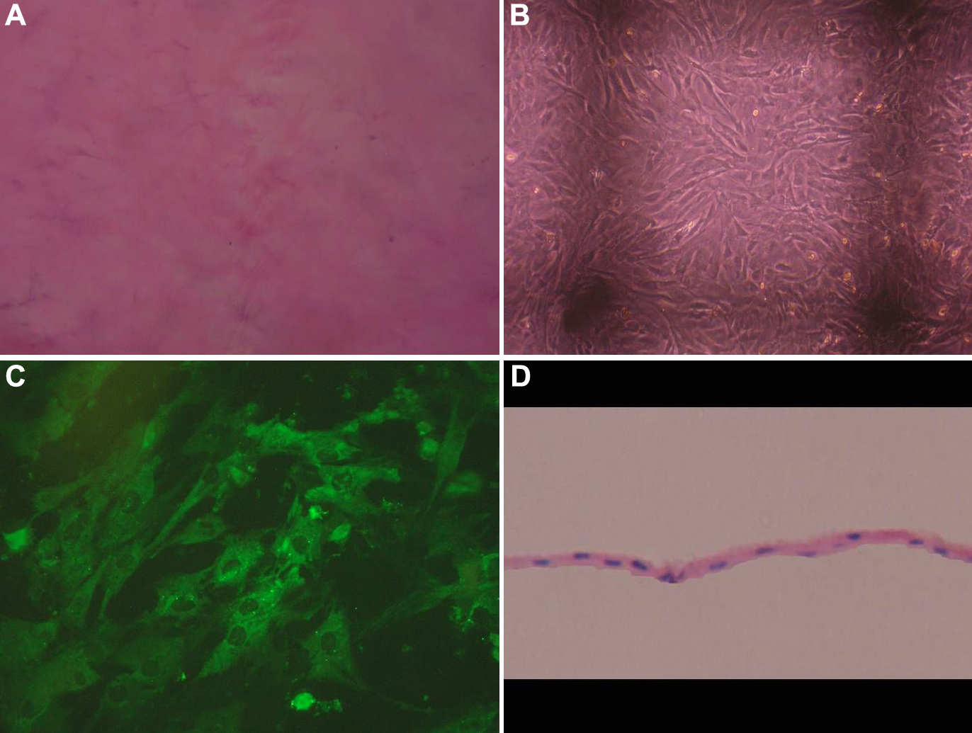
 Figure 3 of Jiang, Mol Vis 2010; 16:1304-1316.