Figure 2. Induced MSCs by corneal stromal
cells. A: Phase contrast micrographs of induced MSCs cultured
on day seven (40×). B: Expression of CK12 on induced MSCs was
positive. C: SEM of induced MSCs showed there were large
numbers of epithelial-like cells that were connected even in a patch
and the structure of the cellular tight junction was clear (arrow;
1000×).
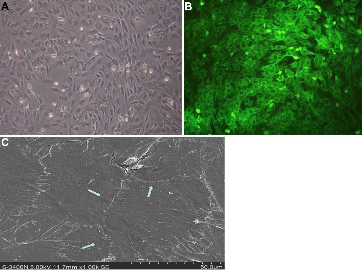
 Figure 2 of Jiang, Mol Vis 2010; 16:1304-1316.