Figure 1. Subcultured mesenchymal stem cells (MSCs) in vitro. A: Phase contrast micrographs of mesenchymal stem cells cultured on day seven (40×). B: Immunofluorescence staining of CD29 was positive. C: Flow cytometric analysis showed that the positive rates of CD29, CD34, CD44, CD45, CD71, CD90, and CD133 were 81.56%, 0.10%,
88.77%, 0.17%, 10.02%, 98.43%, and 1.56%, respectively. D: Scanning electron microscopy (SEM) of MSCs showed a long spindle-shaped appearance with rich processes (1000×).
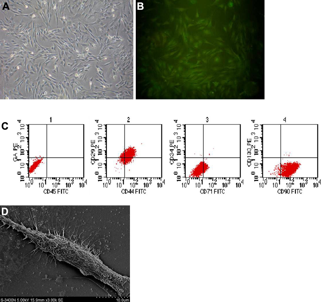
 Figure 1 of
Jiang, Mol Vis 2010; 16:1304-1316.
Figure 1 of
Jiang, Mol Vis 2010; 16:1304-1316.