Figure 3. Cell cycle detection and the
proliferative landscape of whole lenses. A: EdU-positive
(S-phase) cells in the whole lens from an 8-month-old lens. B:
Cells undergoing mitosis are labeled using a fluorescent-conjugated
antibody to phosphorylated-histoneH3 in an 8-month old lens. C:
Total cells in an image field labeled with DRAQ-5. D: Merged
image of all three labels detecting two phases of the cell cycle in the
germinative zone of a whole lens. E: Whole lenses were placed
anterior side down. Image stacks (275 μm) were acquired in the Z-plane
from the center of the anterior pole of the lens, demarcated by the
asterisk. Z-stacks were then projected in the Y-plane and joined
together to re-create the entire lens image. Depth coding was then
applied to each stack to provide relevant distance from the initiation
point of each z-stack. Cooler colors are closer to the asterisk (origin
of the z-stack) than are warmer colors. Using this method, the
germinative zone of an entire lens can be seen in a 1-month old lens. F:
Reconstruction
of an 8-month-old lens showing the decrease in the
number of cells in S-phase with age. G: A larger image of the
boxed area in (E), showing the color scheme of the depth coding
application. H: Larger image of the boxed area in (F).
Scale bars: 10 μm (A-D) and 200 μm (E-H).
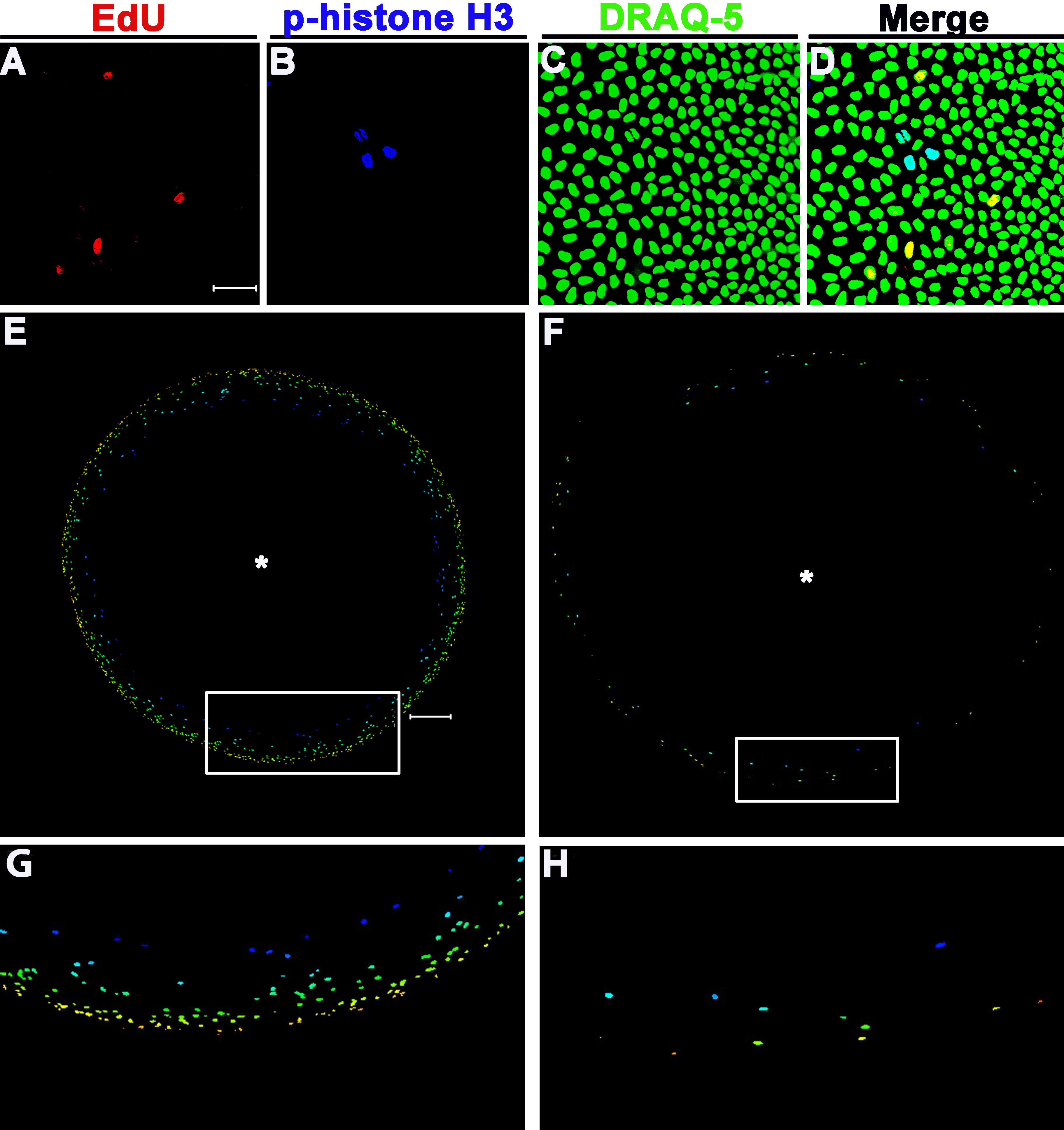
 Figure 3 of Wiley, Mol Vis 2010; 16:1253-1259.