Figure 2. Comparison of whole lenses
labeled with EdU and lens explants labeled with BrdU. A: EdU
labels lens epithelial cells in S-phase in the germinative zone (GZ) of
a 1-month-old lens. Lenses were placed on their equators and image
fields were captured after moving one cells width anteriorly from the
meridional rows (MR) which serve as a geographical marker for the
posterior boundary of the lens epithelium. An example is provided in
the inset, which is stained with Alexa Fluor488 phalloidin to label
filamentous actin. The vertical dashed line is included to roughly
depict the end of the transitional zone (TZ) and the beginning of the
GZ. B: EdU-positive cells in the GZ of an 8-month old lens. C:
An
example
of
labeling of S-phase cells using an antibody to BrdU in an
explant from a 1-month-old lens. Consistent identification of the
germinative zone is complicated by distortion of the explant during
dissection. During dissection, there is unavoidable damage to the lens
epithelium (asterisks). Finally, the explant is difficult to keep
flattened, leaving areas of the explant out of focus and less intense,
making quantification more difficult. D: Lens explant from an
8-month-old mouse labeled with BrdU. E: Quantification of the
percentage of lens cells in S-phase (EdU-positive) from an 8-month-old
Balb/c mouse kept under normoxic conditions (room air; 21% O2)
compared
to
a
mouse breathing 60% O2. The asterisk indicates
a p<0.05). Total cells (green) were stained with the vital dye,
DRAQ-5. Scale bars: 10 μm (A, B) and 50 μm (C, D).
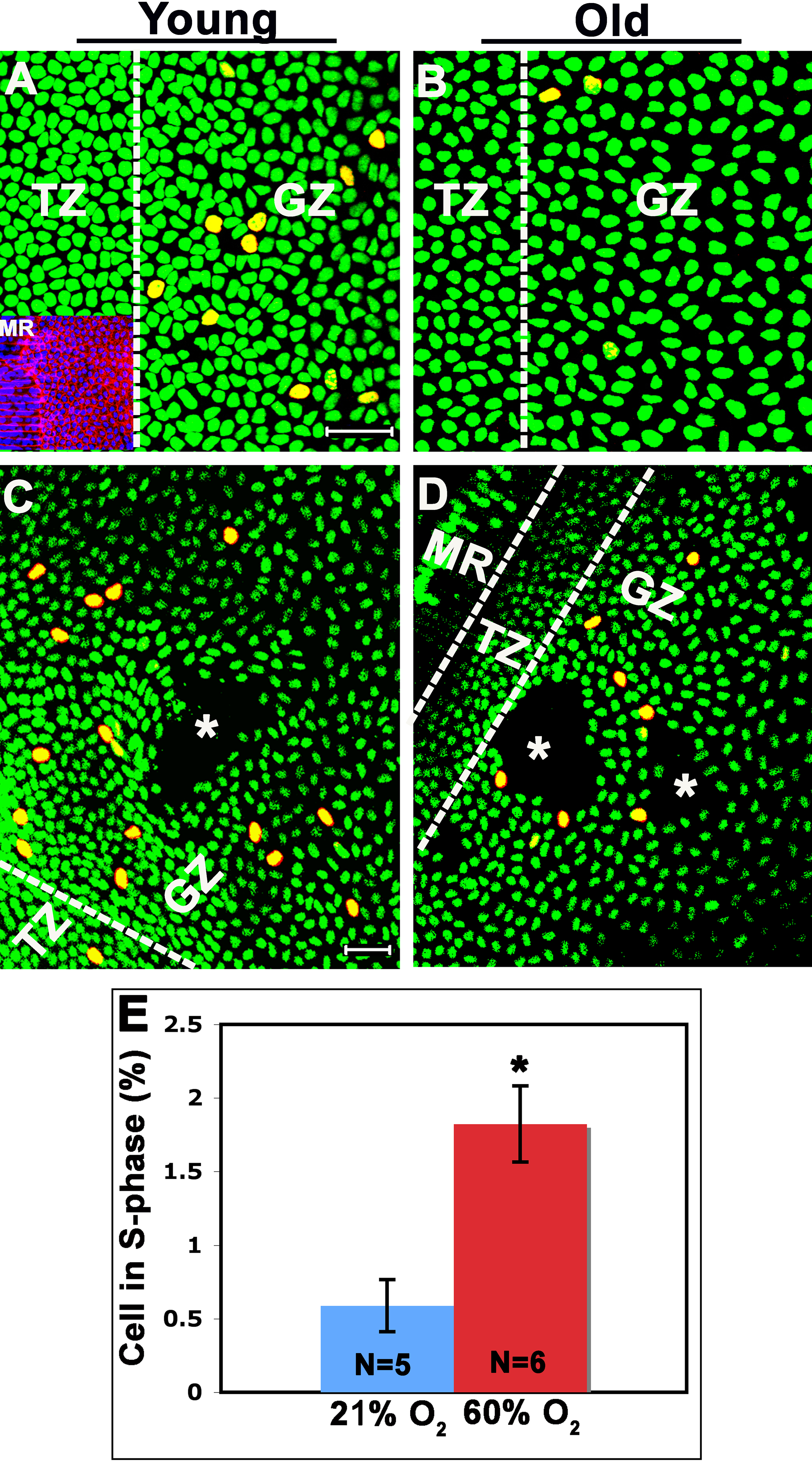
 Figure 2 of Wiley, Mol Vis 2010; 16:1253-1259.