Figure 1. Apparatus used to image whole
lenses. A: Overhead view of the apparatus. Two pieces of rubber
(R) glued to a glass coverslip make a “wedged” well in which to hold
and stabilize the lens (LE) on its equator (dashed line). The coverslip
can then be held in place on the microscope stage and imaged from below
by the microscope objective (Obj). Because the lens is held stationary
in the rubber well, it can gently be rotated using forceps. In this
way, the entire lens equator, including the transitional and
germinative zones can be imaged. B: Side view; the lens is
still in its normal, spherical shape. When rounded, the image plane is
small. C: En face view of the set-up after gently applying a
weighted Petri dish (WPD) on top of the lens. This flattens the fixed
lens, allowing for capture of full image fields.
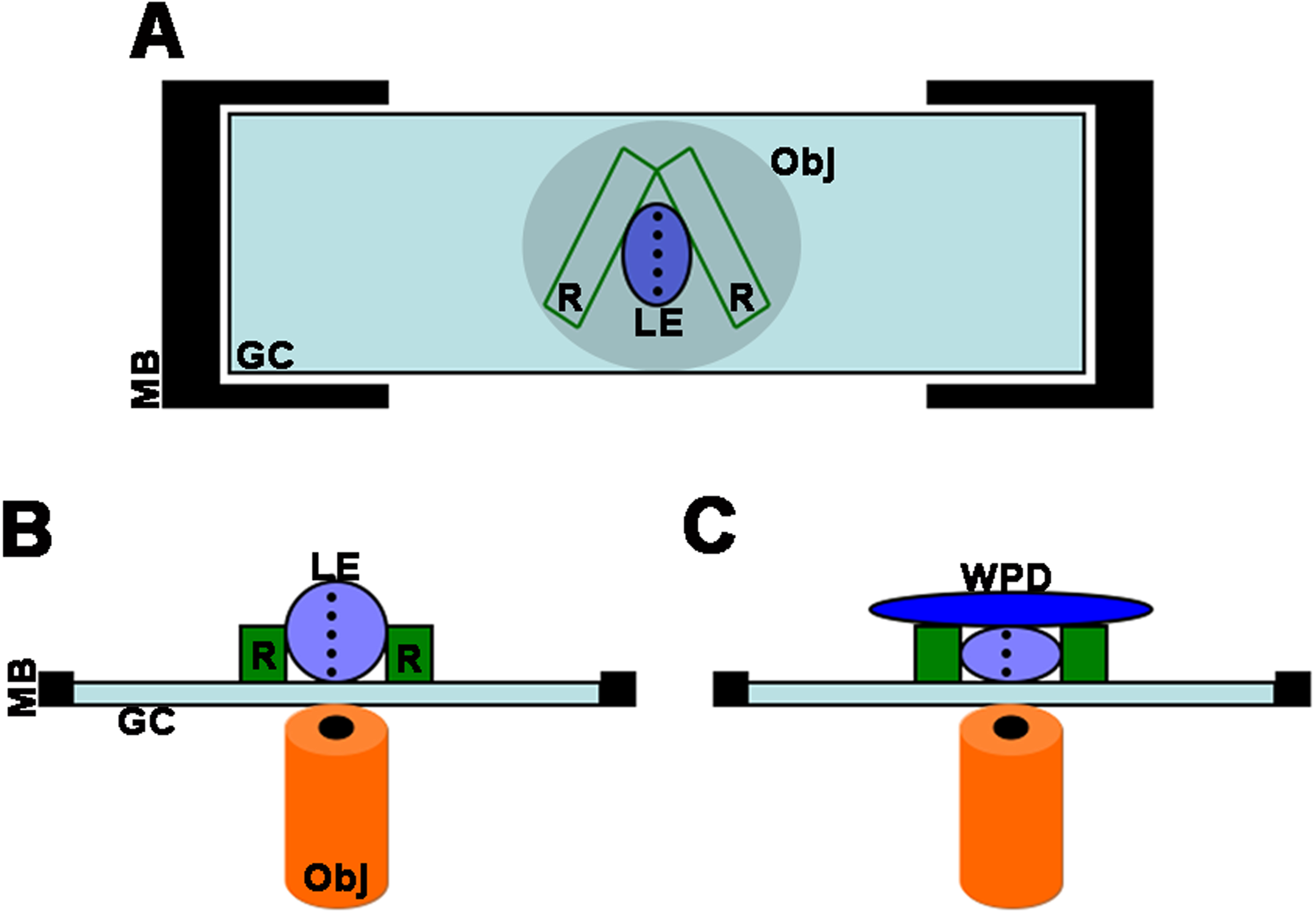
 Figure 1 of Wiley, Mol Vis 2010; 16:1253-1259.