Figure 4. Expression of NPR-A and NPR-B in human p-CEPI cells and CEPI-17-CL4 cells as determined by western blot analysis. A: The presence of NPR-A protein (band observed at 40–55 kDa) in CEPI-17-CL4 and in human p-CEPI cells (from 56, 53, and 56 year old donors) is shown. B: The presence of NPR-B protein (band observed at 24 kDa) is shown for three different donors of human p-CEPI cells (ages 40, 53, and 56) cells and in CEPI-17-CL4 cells. C: The expression of NPR-A and NPR-B in human p-CEPI and CEPI-17-CL4 cells was normalized to GAPDH. The figure shows an apparent lower expression of NPR-A in CEPI-17-CL4
cells than in human p-CEPI cells (p<0.1); the expression of NPR-B was higher in CEPI-17-CL4 than that in human p-CEPI cells (p<0.05). The NPR-B expression was greater than NPR-A expression in CEPI-17-CL4 cells (p<0.05). However, the both
receptor subtypes were expressed to the same extent in the p-CEPI cells (p<0.1).
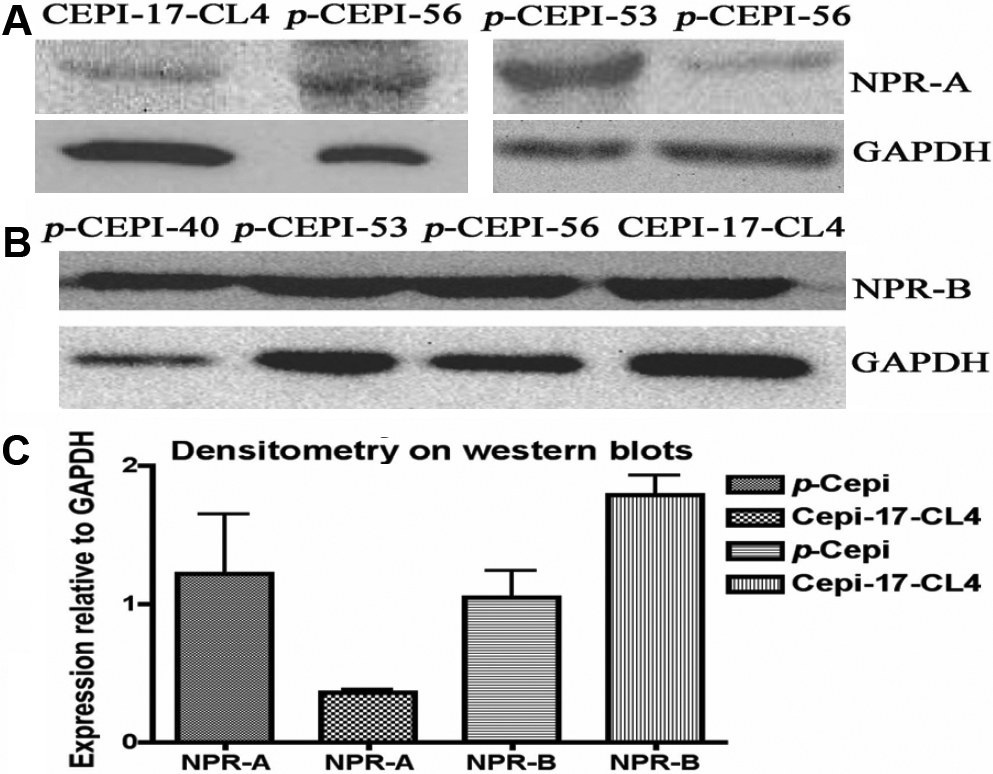
 Figure 4 of
Katoli, Mol Vis 2010; 16:1241-1252.
Figure 4 of
Katoli, Mol Vis 2010; 16:1241-1252.