Figure 3. The expression of NPR-A and NPR-B in formalin-fixed human p-CEPI and CEPI-17-CL4 cells by indirect immunofluorescence is shown. A shows a strong expression of NPR-A in human p-CEPI cells, while B shows expression of NPR-A in CEPI-17-CL4 cells. C shows expression of NPR-B in human p-CEPI cells while D shows expression of NPR-B in CEPI-17-CL4 cells. E and F are DAPI labeled controls (treated with secondary antibody only) for human p-CEPI and CEPI-17-CL4 cells, respectively. Each panel is at 40× magnification.
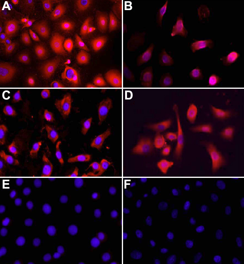
 Figure 3 of
Katoli, Mol Vis 2010; 16:1241-1252.
Figure 3 of
Katoli, Mol Vis 2010; 16:1241-1252.