Figure 2. Expression of NPR-A and NPR-B in paraffin sections of formalin-fixed human corneas as determined by indirect immunofluorescence.
A, B: The presence of NPR-A in the epithelium of the central cornea. A shows stronger expression in the superficial layers when compared with the control (B). C, D: C shows the presence of NPR-A primarily in the superficial layers of the limbal epithelium. The control shows no expression
(D). E, F: E shows strong expression of NPR-B primarily in the superficial cell layers of the central cornea compared when compared with
the control (F). G, H: G shows that NPR-B is very distinctly confined to the superficial epithelium of the limbus compared with the control (H). All panels are at 10× magnification.
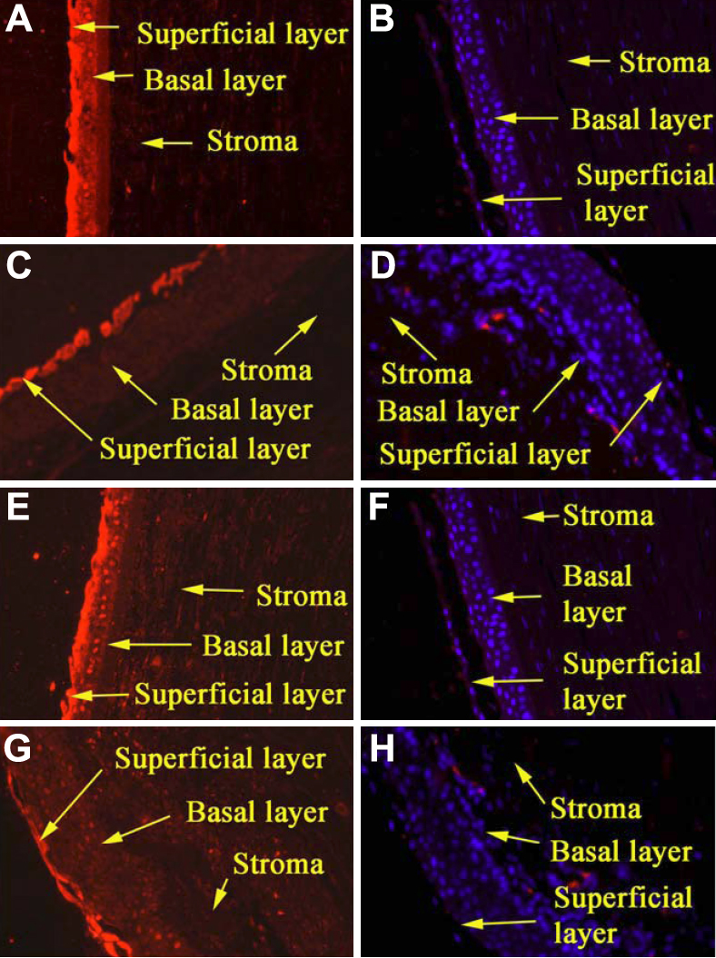
 Figure 2 of
Katoli, Mol Vis 2010; 16:1241-1252.
Figure 2 of
Katoli, Mol Vis 2010; 16:1241-1252.