Figure 2. Characterization of limbal
explant culture derived epithelial (LEC) and mesenchymal like cells
(MC-L). Following immunoctytochemical analysis (A-L)
epithelial cells showing positive for ABCG2 (A), CK3/CK12 (B;
green
fluorescence), CK19 (C; red fluorescence), CK14 (D),
E-Cadherin (E), vimentin (F; green fluroscence), double
immunostaining for PAX-6 (green fluorescence) and vimentin (red
fluorescence; G). Mesenchymal like cells of limbus (H-L)
showing
positive for vimentin (H) and negative for cytokeratin
3/12 (I), cytokeratin 14(J), CD34 (K), and nestin (L).
Nuclear
staining was performed with propidium iodide (red; A, B,
D, E, H-L). Scale bar=20 µm (A-C,
E-H, and J) and 10µm (D, I, K,
L). Flow cytometry analysis (M-V) was performed by
incubation of the mesenchymal like cells of limbus with the indicated
antibodies. M: Isotype controls for FITC and PE, N:
CD90 FITC and CD44 PE, O: CD40-FITC and HLA-ABC PE, P:
CD11b FITC and HLA-ABC PE, Q: CD34 FITC and CD10 PE R:
Isotype control for FITC and APC, S: CXCR4 APC and CD90 FITC, T:
CD40L
APC and CD40 FITC, U: Isotype control for FITC and PerCP,
V: CD138 PerCP and CD13 FITC.
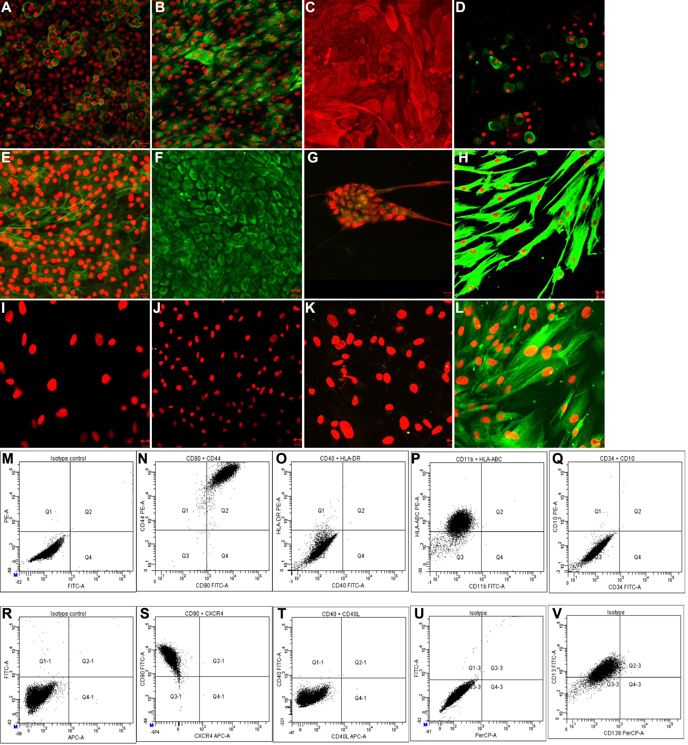
 Figure 2 of Polisetti, Mol Vis 2010; 16:1227-1240.