Figure 1. Morphological features of
mesenchymal cells of limbus. Limbal explant cultures having epithelial
(E) and mesenchymal cells (S; 200×; A). Cell sphere formation
in the MC-L cultures giving impression of embryoid body formation
(200×; B). Spindle shaped morphology of MC-L forming colonies
(200×; C). Culture of MC-L showing both spindle shaped and
broad flattened cells (arrows; 200×; D)
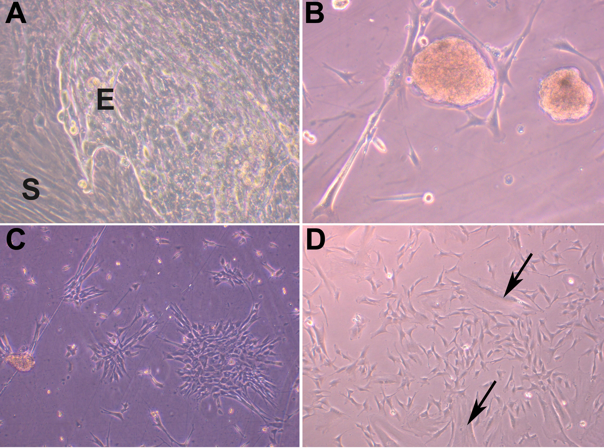
 Figure 1 of Polisetti, Mol Vis 2010; 16:1227-1240.