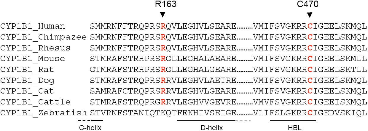
Figure 3. Multiple amino-acid sequence
alignment of CYP1B1 from different species. Sequence alignment was
generated by
ClustalW.
The
positions of mutated amino-acids newly reported in this study are
indicated by arrows and red letters. The COOH-terminal amino-acids of
C-helix, NH
2-terminal amino-acids of D-helix and heme
binding loop (HBL) are indicated below the sequence alignment, by a
line.
 Figure 3 of Hilal, Mol Vis 2010; 16:1215-1226.