Figure 8. Expression patterns of phospho-adducin in the lens epithelia of injured lenses. At day 2, many lens epithelial cells were
labeled for phospho-adducin in an injured wild-type (WT) lens (A, asterisk). On the other hand, in a tenascin-C null (KO) injured lens the epithelial cells at the leading edge were not labeled
for phospho-adducin (B). At day 5, many epithelial cells were positive for phospho-adducin in the multicellular layer at the capsular break site
in a WT mouse (C, asterisk), although its immunoreactivity was weaker than that in the cells at day 2, whereas phospho-adducin was only faintly
detected in the majority of epithelial cells (star) in an injured KO lens at day 5 (D). At day 10, the cells around the break in the anterior capsule were not labeled for phospho-adducin in either WT (E) or KO (F) mice; Bar, 10 μm.
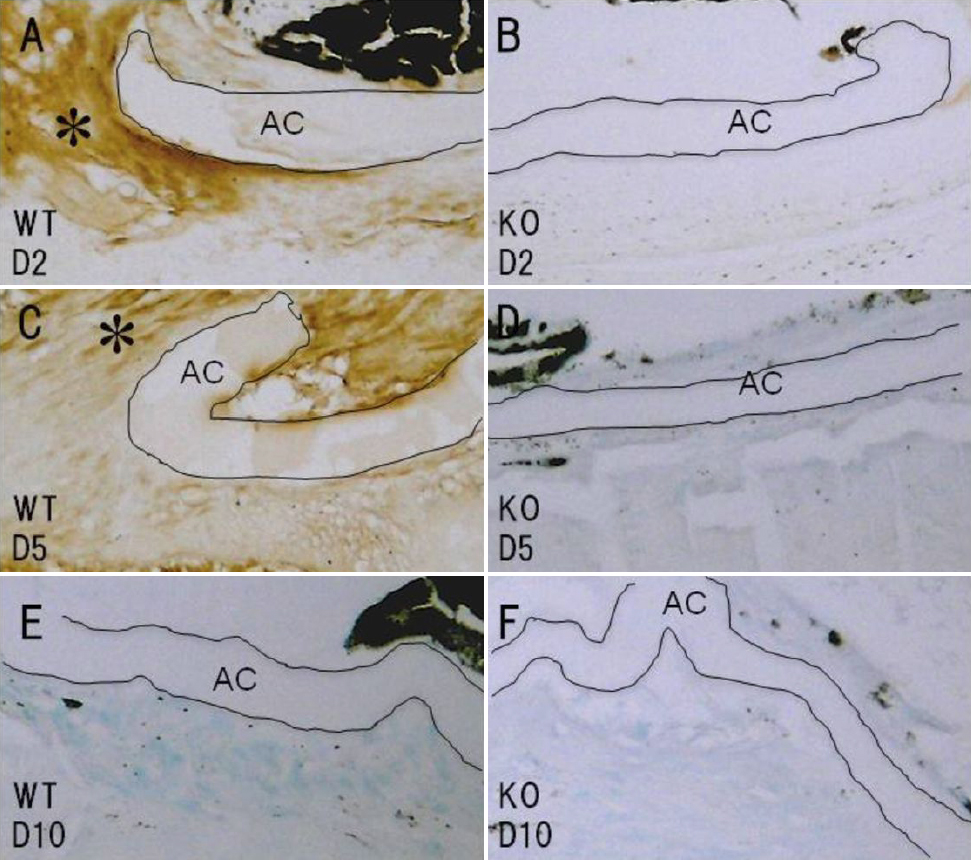
 Figure 8 of
Tanaka, Mol Vis 2010; 16:1194-1205.
Figure 8 of
Tanaka, Mol Vis 2010; 16:1194-1205.