Figure 7. Expression patterns of phospho-Smad2 in the lens epithelia of injured lenses. Uninjured lens epithelial cells beneath the
anterior capsule were not labeled for phosph-Smad2 in either the wild-type (WT; A) or tenascin-C null (KO; B) mice. At day 2, the nuclei of some epithelial cells were labeled for phospho-Smad2 in an injured WT lens (C, arrows). On the other hand, in a KO injured lens the nuclei of the epithelial cells at the leading edge of the cellular
multi-layer were not labeled for phospho-Smad2 (D). At day 5, many epithelial cells were founded to contain nuclei that were positive for phospho-Smad2 in the multicellular
layer at the capsular break site in a WT mouse (E, arrows), whereas no phospho-Smad2 was detected in the majority of epithelial cells in multicellular layer in an injured
KO lens (F). At day 10, the cells around the break in the anterior capsule were not labeled for phospho-Smad2 in either the WT (G) or KO (H) mice. Dotted lines, border between the multilayered cells and the lens cortex, AC, anterior capsule; Bar, 20 μm.
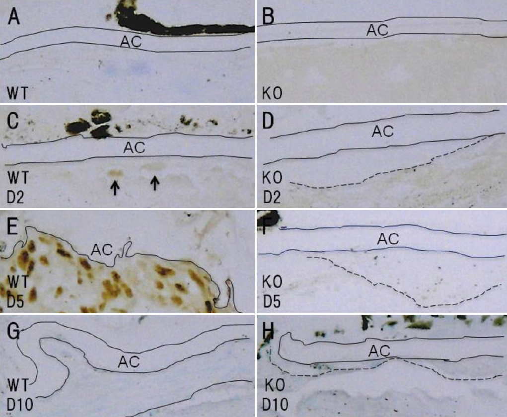
 Figure 7 of
Tanaka, Mol Vis 2010; 16:1194-1205.
Figure 7 of
Tanaka, Mol Vis 2010; 16:1194-1205.