Figure 2. Histology of the edge of the break in the anterior lens capsule in wild-type (WT) and tenascin-C null (KO) mice. At day 2,
the lens epithelial cells around the capsular break (arrows) exhibited an epithelial-like morphology in both the WT (A and insert; asterisk) and KO (B and insert; asterisk) mice. At day 5, the epithelial cells had formed a multilayer at the edge of the broken capsule in both
the WT (C) and KO (D) mice. The wound indicated by the arrows was filled with cells in the WT mice (C), while that in the KO mouse lens remained open (D). At a higher magnification, the WT cells (C, insert) showed an elongated fibroblast-like shape (asterisk), while those of the KO lens (D, insert) exhibited an epithelial-like morphology (asterisk). At day 10, a multi-cellular layer had formed beneath the anterior
capsule in both the WT (E) and KO (F) mice. At a higher magnification, the cells in both groups (E and F; inserts) exhibited an elongated fibroblast-like shape. Inserts of A to F show the higher magnification images of the boxed areas in A through F. AC, anterior lens capsule, Bar, 10 μm (A through F), or 25 μm for each of the inserts.
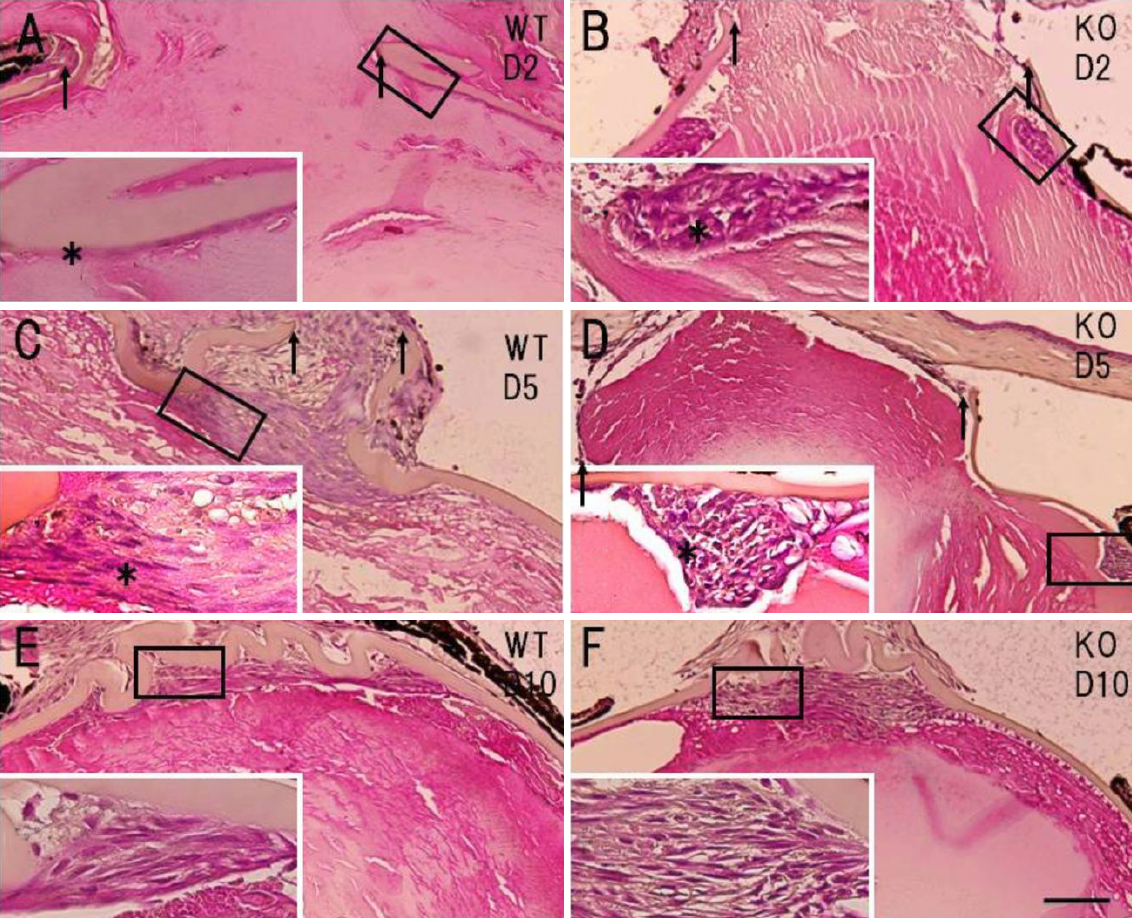
 Figure 2 of
Tanaka, Mol Vis 2010; 16:1194-1205.
Figure 2 of
Tanaka, Mol Vis 2010; 16:1194-1205.