Figure 4. Light-induced signal
transmission to the inner retina. After complete dark adaptation,
animals and organ cultures were exposed to 1 Hz stroboscopic
illumination for 2 h. This procedure examines whether the inner retina
can respond to a sustained signal from the photoreceptors. A:
C-fos expression was induced by stroboscopic illumination in cells in
the inner nuclear layer and the retinal ganglion cell layer of the
intact animal. D: In organ cultures, c-fos expression could
also be demonstrated using this paradigm. Images are representative
examples from organ cultures derived from three different litters. For
each image, a corresponding bright-field image is provided for
orientation (B and C). In the absence of stroboscopic
illumination, no c-fos immunoreactivity was observed in vivo (E)
or in vitro (F). G: A no-primary antibody control is
provided for nonspecific staining of the secondary antibody.
Abbreviations: GCL, ganglion cell layer; INL, inner nuclear layer; IPL,
inner plexiform layer; IS, inner segments; ONL, outer nuclear layer;
OPL, outer plexiform layer; OS, outer segments. The scale bar
represents 20 μm.
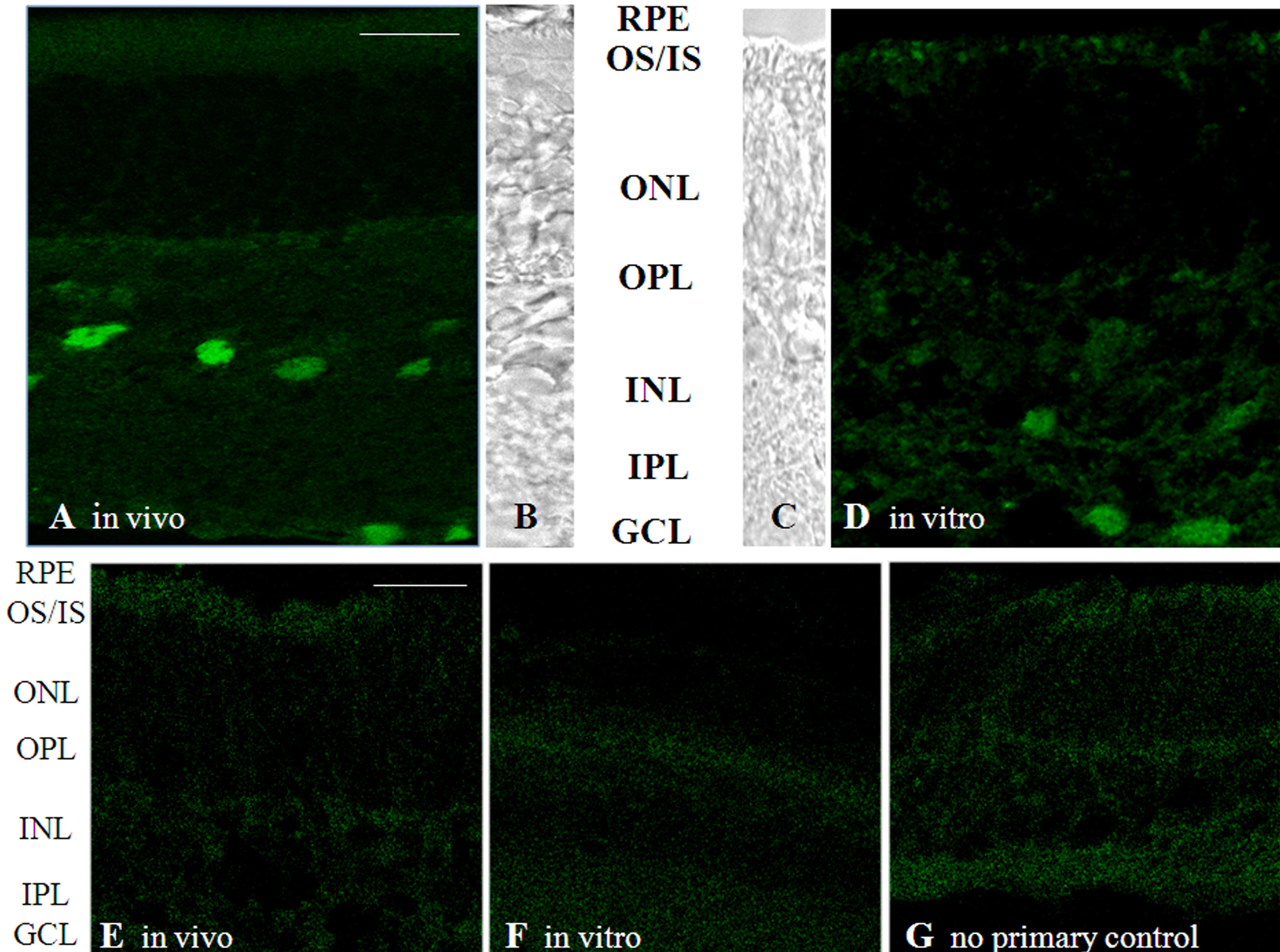
 Figure 4 of Bandyopadhyay, Mol Vis 2010; 16:1178-1185.