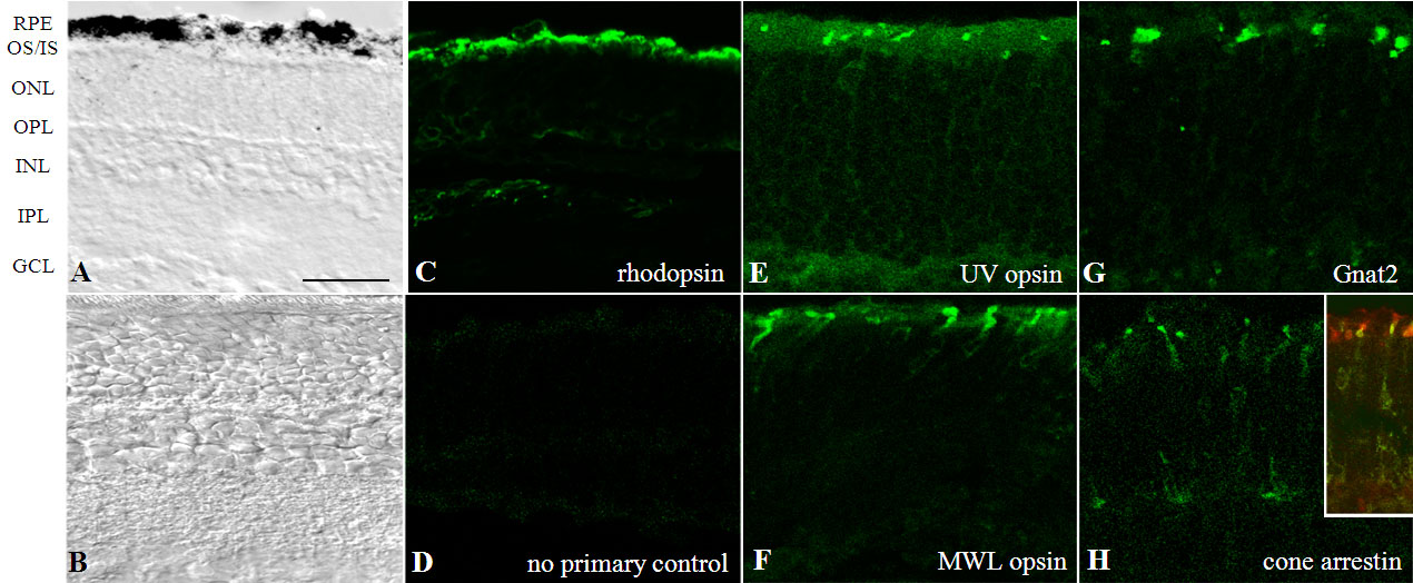
Figure 1. Establishment of a mouse
retina–retinal pigment epithelium (RPE) explant system.
A: The
bright-field micrograph of a C57BL/6 mouse retinal explant placed in
culture at postnatal day 7 (P7; which is called days in vitro 0, DIV0)
and analyzed at DIV11 (P18=P7+11DIV) shows that normal retinal layers
are formed and maintained in situ.
B: A P18 C57BL/6 mouse
retina is provided for comparison. In culture, rhodopsin (
C),
ultraviolet (UV) opsin (
E), middle-wavelength (MWL) opsin (
F),
and
cone transducin (
G) are trafficked properly to the
photoreceptor outer segment.
H: Cone arrestin is present in the
inner segments and cone pedicles, as described for light-adapted
retinas [
30]; a
double labeling (inset of panel
H) of cone arrestin with
PNA-lectin is provided to allow for visualization of inner and outer
segments.
D: No-primary antibody control is provided for
nonspecific staining of the secondary antibodies. Images are
representative examples from three to five different litters.
Abbreviations: GCL, ganglion cell layer; INL, inner nuclear layer; IPL,
inner plexiform layer; IS, inner segments; ONL, outer nuclear layer;
OPL, outer plexiform layer; OS, outer segments. Scale bar 50 μm.
 Figure 1 of Bandyopadhyay, Mol Vis 2010; 16:1178-1185.