Figure 2. Images of human primary lens epithelial cells infected with different lentivirus. A, C, E, G, and I were taken under bright field. B, D, F, H, and J were taken under green fluorescent protein (GFP) field. A and B show the control group; the human primary lens epithelial cells were not infected in this group. C and D show group1 (mock group) infected with shRNA-scramble (random siRNA sequence). E and F show group 2, which is infected with shRNA1. G and H show group 3, which is infected with shRNA2. I and J show group 4, which is infected with shRNA3. The control group (B) has no GFP fluorescence, while almost 80–90% of the cells of group1, 2, 3, and 4 have green fluorescence.
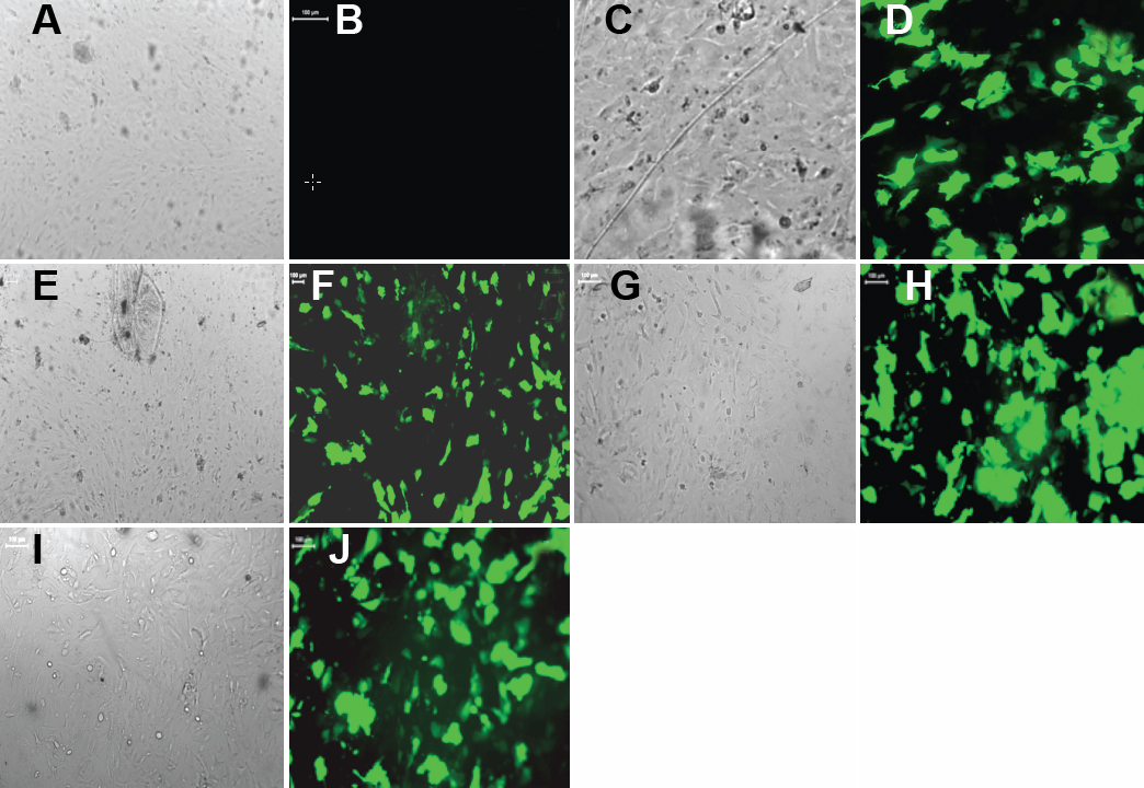
 Figure 2 of
Yang, Mol Vis 2010; 16:105-112.
Figure 2 of
Yang, Mol Vis 2010; 16:105-112.