Figure 4. Retrospective analysis of the patients with sclerocornea for further ocular abnormalities. A: Axial and sagittal T2-weighted MRI scans of the head and orbits of two affected members 1966 and 1967 (aged 25 and 22 years
old) from the MEP54 pedigree. Both patients demonstrate aphakia as depicted by the absence of a dark lens in the anterior
part of the eye. In the absence of surgery, the left eye of patient 1966 seems to be phthisical. The axial lengths for 1966
are 10 and 17 mm and 1967 are 19 and 16 mm for the right and left eyes, respectively confirming that there is also microphthalmia.
The sagittal section shows that there are no obvious structural abnormalities of the brain. B: Left eye ultrasound scan of patient 1654 from the Mexican pedigree showing an optic disc coloboma (white arrow).
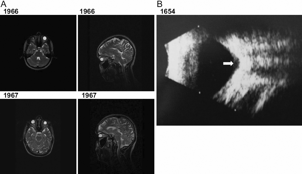
 Figure 4 of
Ali, Mol Vis 2010; 16:1162-1168.
Figure 4 of
Ali, Mol Vis 2010; 16:1162-1168.