Figure 1. Clinical description of the families. A: The pedigree structures are shown for MEP54 and the Mexican pedigree. Affected individuals are depicted with filled-in symbols.
The numbers highlight the family members from whom blood was taken for DNA extraction. B: Anterior segment photos were taken using a Nixon Camera for affected member 1971 (aged 8 years) from the Pakistani and 1855
(aged 12 years), 1652 (aged 40 years), 1655 (aged 38 years) and 1851 (aged 48 years) from the Mexican pedigrees. Note total
sclerocornea. Acuity was hand movements only.
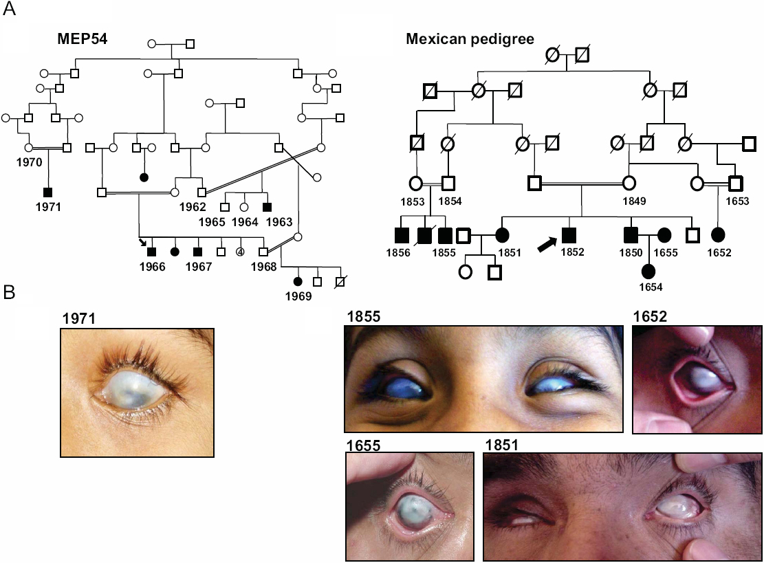
 Figure 1 of
Ali, Mol Vis 2010; 16:1162-1168.
Figure 1 of
Ali, Mol Vis 2010; 16:1162-1168.