Figure 5. Teratoma formation examination
and alkaline phosphatase (AKP) staining. A: HE staining shows
teratoma from ES cells and P9 (wk 4) e-Pc containing multiple tissues,
including epithelium (E), neural (N), muscle (M), and glandular
structures (G). The scale bar represents 50 μm. B: AKP
staining in P9 (wk 4) and P18 (wk 8) e-Pc (magnification, 100×). AKP
staining was positive at P9 (week 4), and positive but weaker at P18
(week 8). Small and cohesive colonies were mostly observed in P9 cells.
Flatter, larger and more migratory colonies were noted in P18 cells.
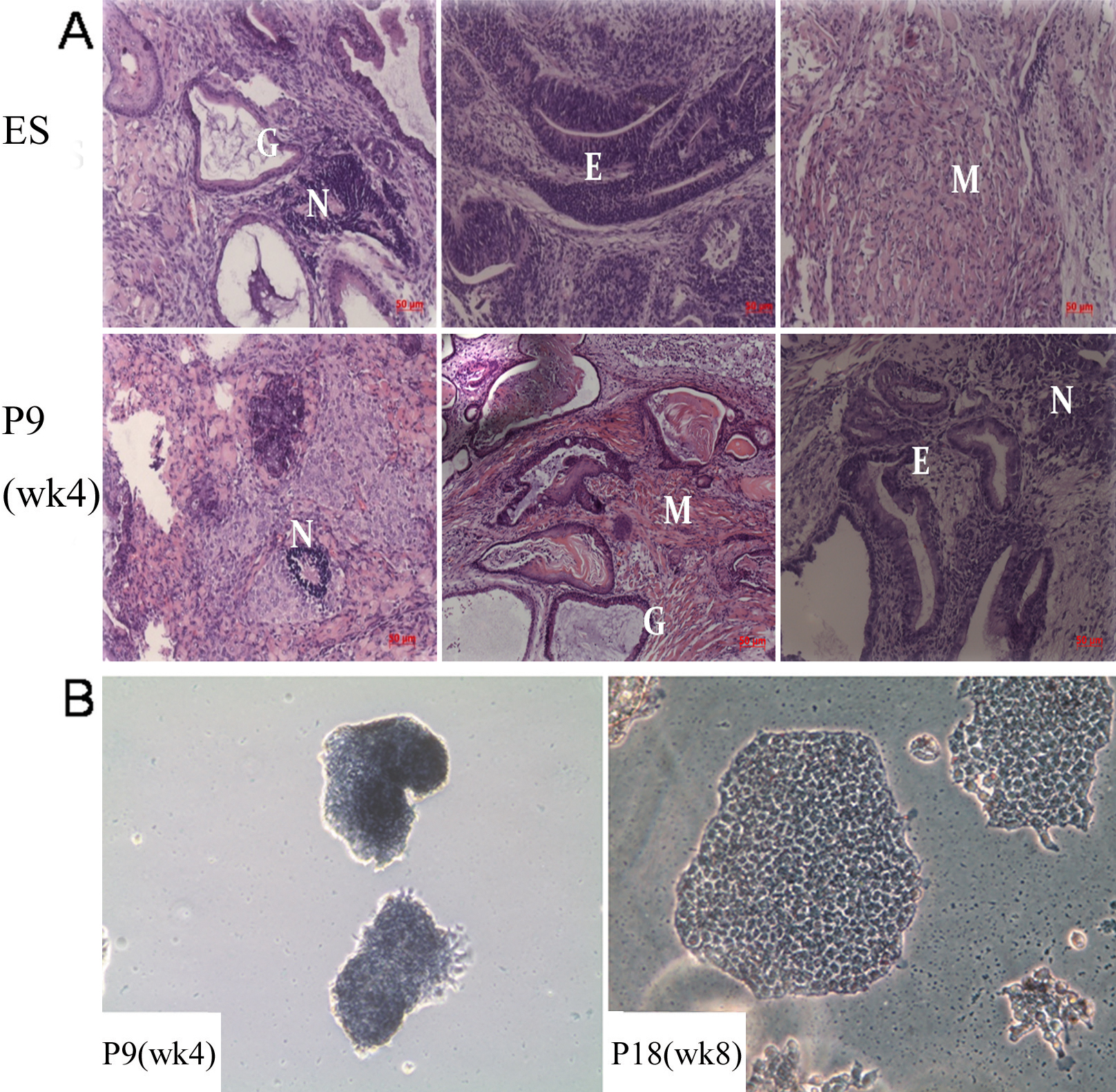
 Figure 5 of Zhan, Mol Vis 2010; 16:1154-1161.