Figure 4. Expression of corneal-epithelium-related proteins in different conditions and passages with immunofluorescent staining. A-C: p63, ABCG2, and K3 (green, positive cells; blue, nuclei) for P2 in all groups and P9, P18 of e-Pc. D: Vimentin for P6 in p-Pc group. E: Vimentin for P9 in e-Pc group. The scale bar in B represents 50 μm. Corneal tissue-specific marker K3 and progenitor cell markers, p63 or/and ABCG2 were still found in different
passages of e-Pc. Vimentin, an intermediate filament protein and a characteristic of keratocytes and fibroblasts was not detected
in P9 cells of e-Pc. But it was positive in P6 of p-Pc.
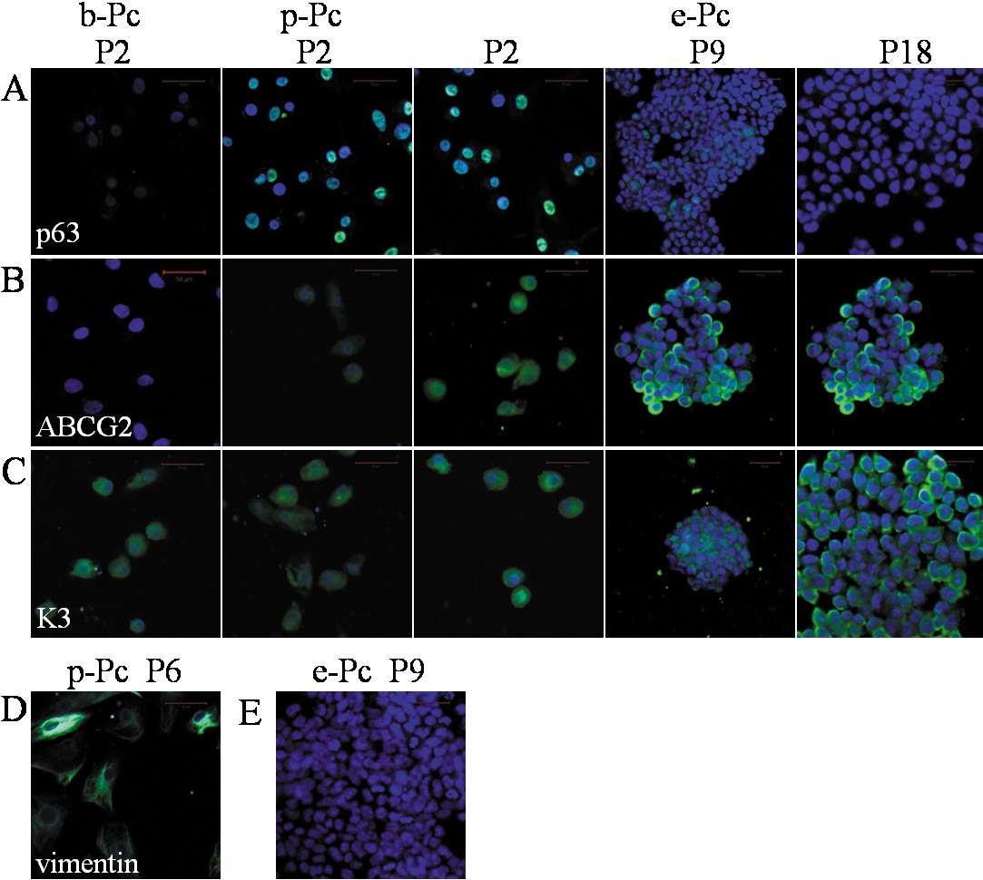
 Figure 4 of
Zhan, Mol Vis 2010; 16:1154-1161.
Figure 4 of
Zhan, Mol Vis 2010; 16:1154-1161.