Figure 2. Photographs of fundi from WS1 patients with PAX3 mutations. Fundus photos were taken from the right (OD) and the left (OS) eyes of two patients, II:1 from family C and II:1
from family E. The colors of fundus photos were different between two eyes in both patients, where mild retinal hypopigmentation
was demonstrated in the left eyes of both patients. The difference of fundus colors between C_II:1 and E_II:1 is of no clinical
significance as different fundus cameras were used. Except for hypopigmentation, the fundus structure was comparatively normal
in the patients.
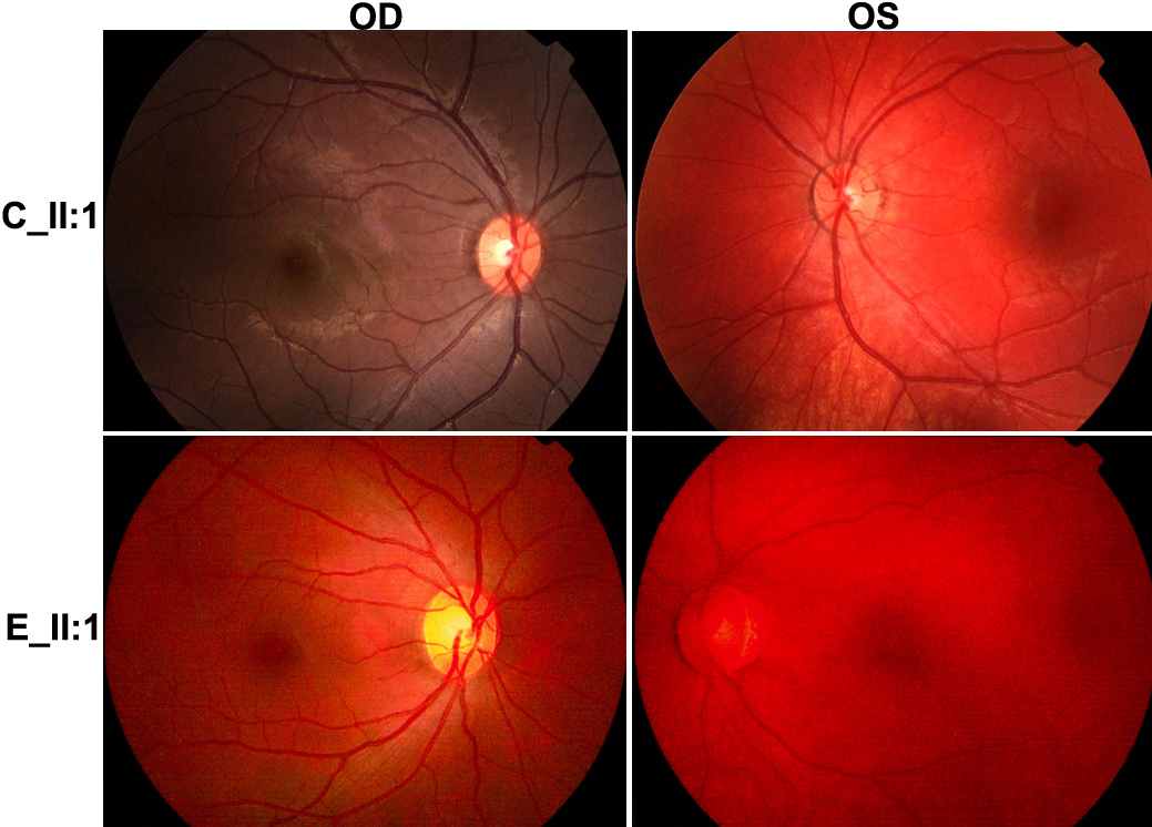
 Figure 2 of
Wang, Mol Vis 2010; 16:1146-1153.
Figure 2 of
Wang, Mol Vis 2010; 16:1146-1153.