Figure 4. Sequence of the PCR product of
exon 5 in PAX6. The top chromatogram represents the sequence of
a normal family member (normal). The middle chromatogram shows a
reading frame shift in the proband (III-25, heterozygous), and the
arrow indicates the initiation of the mutation site (beginning of
overlapping peaks). The bottom chromatogram exhibits the sequence of
the extra band of the SSCP removed from the gel (mutant), and the arrow
indicates the location of the mutation.
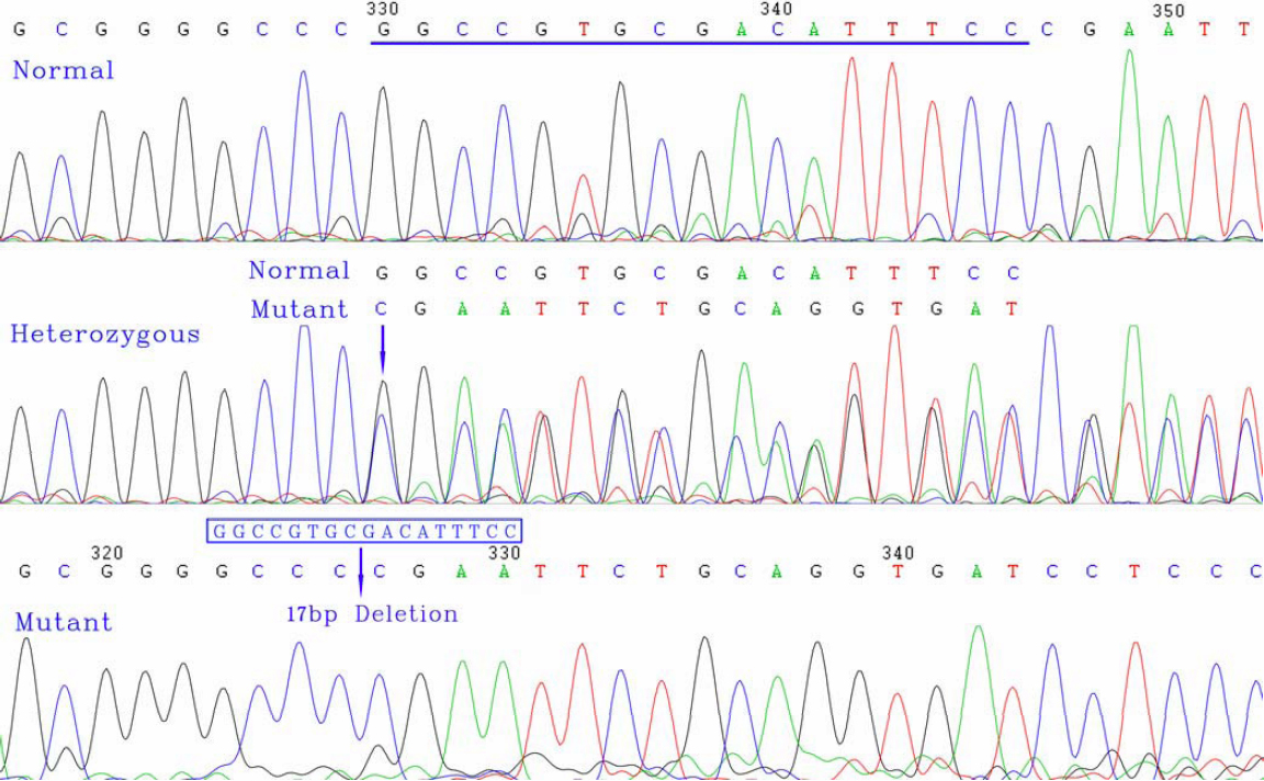
 Figure 4 of Cai, Mol Vis 2010; 16:1141-1145.