Figure 6. ORO staining for lipid during
meibomian gland development at different time points. Consistent with
PPARγ, ORO staining was absent at E18.5 (A) and P0 (B).
Lipid production was first detected at P3 (C) within the central
region of the developing duct. At P5 (D) and P8 (E), ORO
staining continued to be limited to the inner core of the developing
duct. After eyelid opening at P15 (F), ORO was present in the
acini of the mature gland.
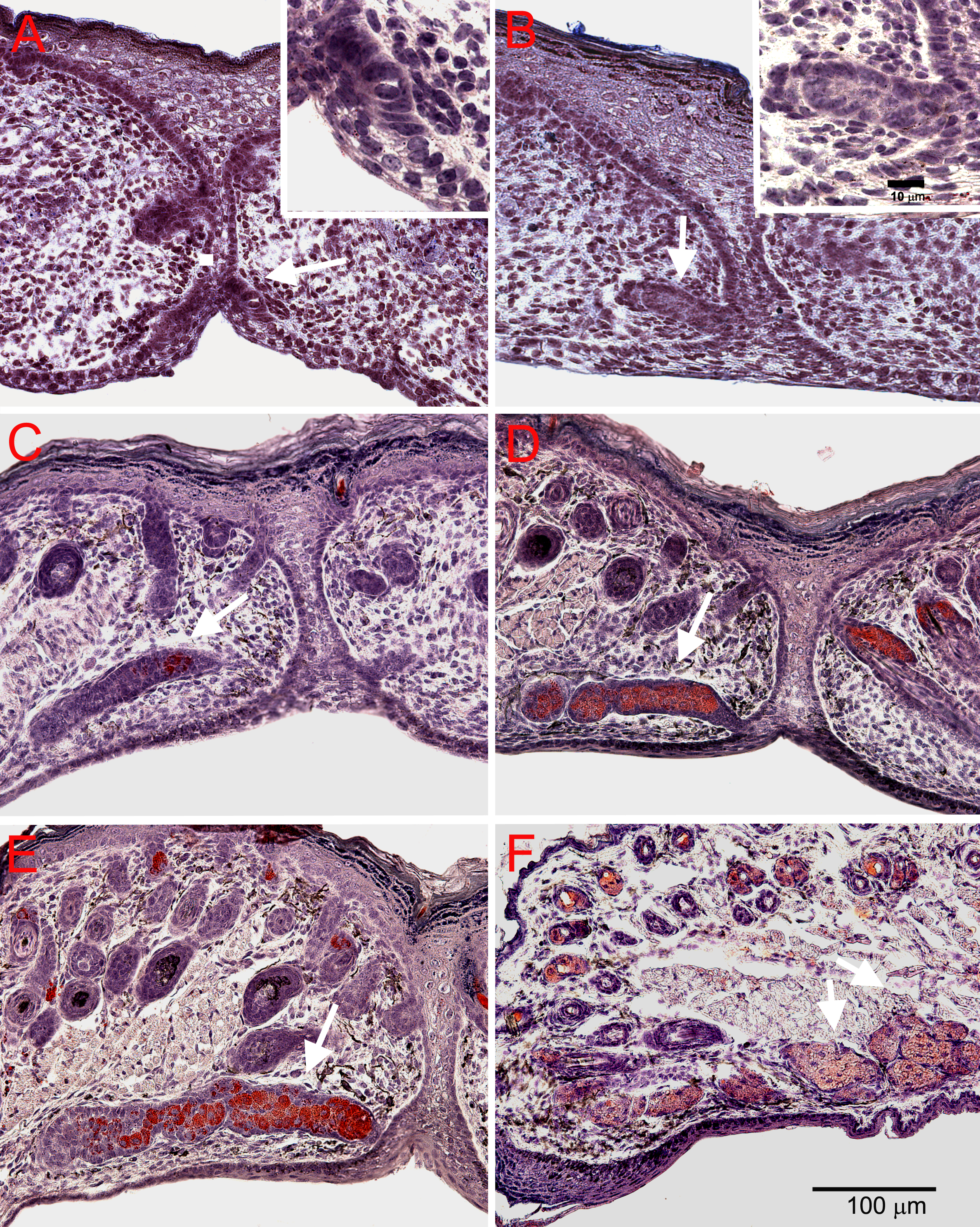
 Figure 6 of Nien, Mol Vis 2010; 16:1132-1140.