Figure 4. PPARγ (Green) and nuclei (DAPI,
red) staining of meibomian glands, white fat, and brown fat from adult,
P60 mice. In the meibomian glands (A), nuclear and extra-nuclear
staining of the plasma membrane and intracellular structures, possibly
microsomes were detected. Staining of white fat and brown fat (B
and C, respectively) showed predominantly nuclear staining.
Western blotting of meibomian gland protein with PPARγ antibodies
identified a 50 kDa protein band (D, lane 1) that was also
present in extracts from white fat and brown fat (D, lane 2 and
3, respectively). Note that the meibomian gland, white fat and brown
fat all showed a second band with a molecular weight of 72 kDa
that may represent post-translational modifications of PPARγ.
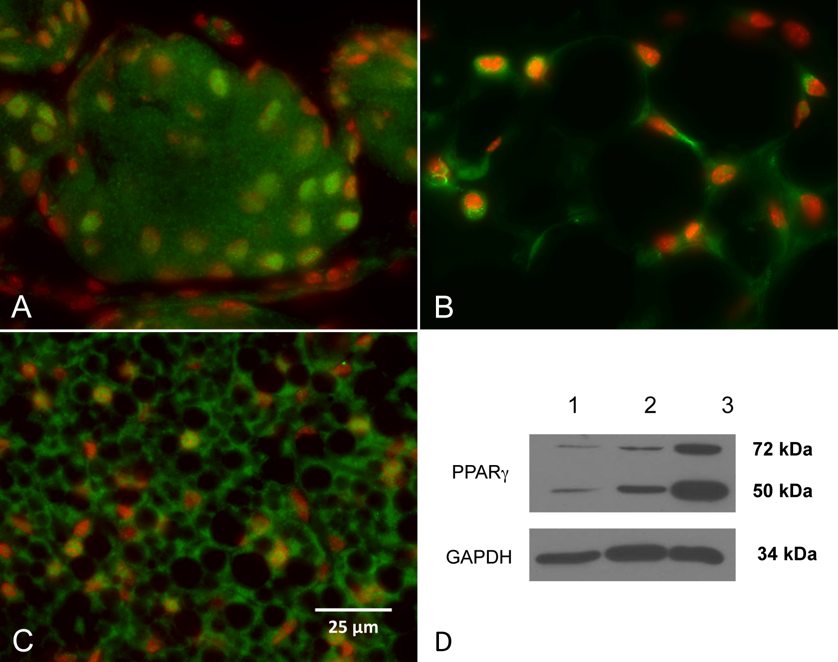
 Figure 4 of Nien, Mol Vis 2010; 16:1132-1140.