Figure 3. Confocal images using actin (red) and nuclei (DAPI, cyan) staining of eyelids at P8 and P15. At P8 (A), developing meibomian glands showed distinct differentiation into ductal and acinar regions. Within the ductal region, basal
epithelial cells along with a single layer of suprabasal cells appear to line a more central region containing enlarged epithelial
cells that lacked actin staining (asterisk). Within the acinar region, the developing meibomian gland duct branched into multiple
ductules that appeared to lack any cellular contents (arrows) and terminated into developing acini. At P15 (B), the meibomian glands appear to obtain an adult meibomian gland morphology.
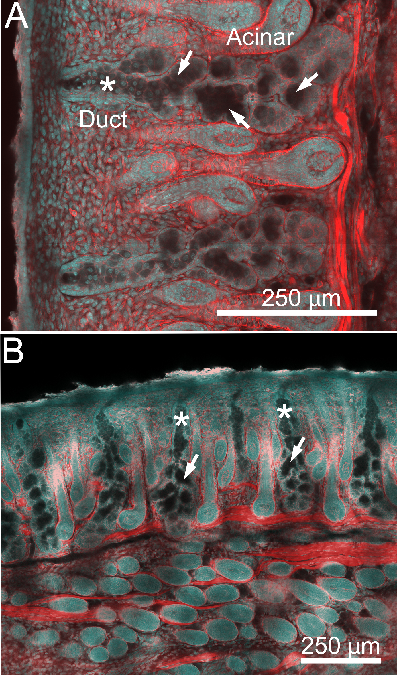
 Figure 3 of
Nien, Mol Vis 2010; 16:1132-1140.
Figure 3 of
Nien, Mol Vis 2010; 16:1132-1140.