Figure 2. Nuclei staining (DAPI) at P3 and P5. At P3 (A and B), meibomian glands appeared to be comprised of a cord of epithelium with a bordering basal cell layer and a two-three cell
suprabasal cell layer within the inner core at the distal end of the developing meibomian gland (A, arrow). At the proximal end there appeared to be a reduction of the number of cells within the inner core of the developing
duct (B, arrow). By P5 (C and D), early branching of the invaginating epithelial cord was observed (arrows). Additionally, there appeared to be enlargement
of cells within the central cord of the epithelium (asterisk).
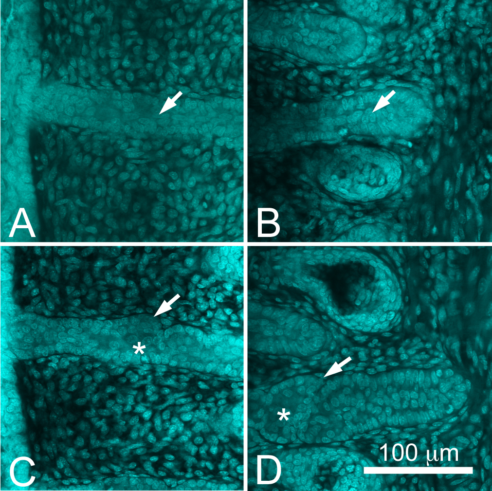
 Figure 2 of
Nien, Mol Vis 2010; 16:1132-1140.
Figure 2 of
Nien, Mol Vis 2010; 16:1132-1140.