Figure 1. Confocal images using actin
(red) and nuclei (DAPI, cyan) staining of eyelids at different time
points of meibomian gland morphogenesis. At E18.5 (A), we
observed the formation of an epithelial condensation (asterisk) within
the fused lid margin. Epithelial placodes in the superior and inferior
lids also appeared opposite to each other, and were associated with
condensation of mesenchyme as detected by cell alignment and increased
actin staining of mesenchyme directly adjacent to the placode (arrows).
At P0 (B), we observed an epidermal invagination (arrow) that
undergoes proliferation and maturation with progressive enlargement and
elongation adopting a tubular shape at P1 and P3 (C and D,
respectively).
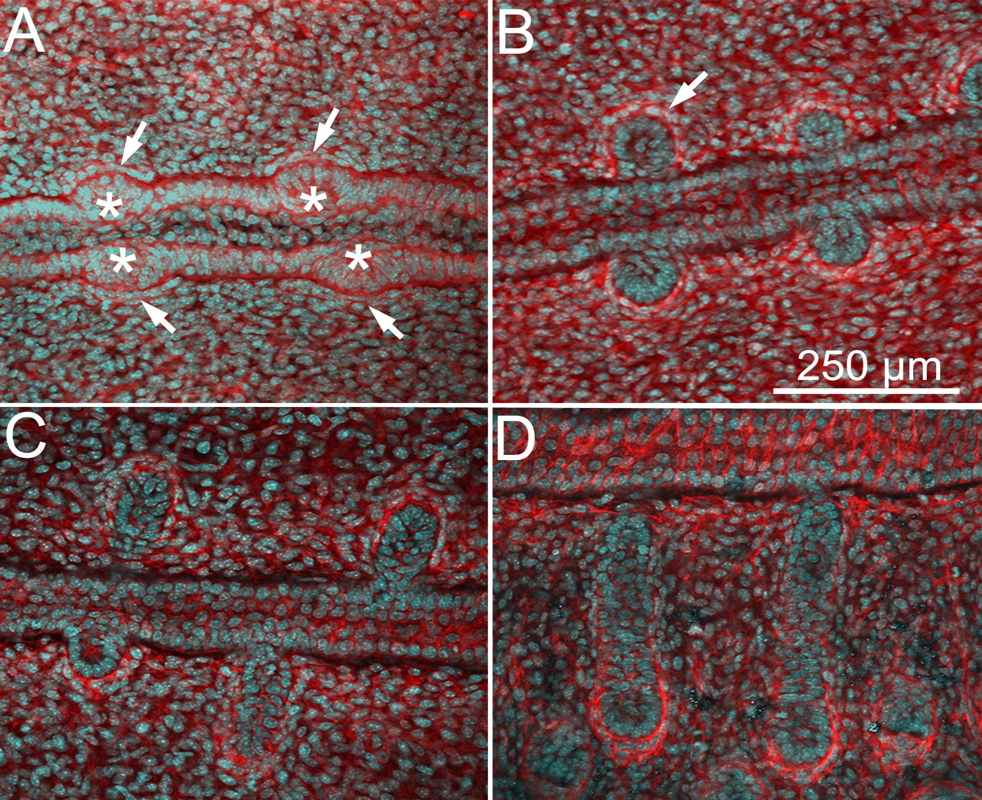
 Figure 1 of Nien, Mol Vis 2010; 16:1132-1140.