Figure 9. Autofluorescence of human retinal pigment epithelium (RPE). Autofluorescence detection of retinal pigment epithelium cells
in the human eye by laser scanning cytometry. A section of the retina from a 14-month-old human male was processed for immunolabeling
up to the primary antibody step. The background fluorescence in each channel was then analyzed. Panel A shows autofluorescence in the green channel. Panel B shows an X–Y position plot of the same data. Most autofluorescent cells fall in a line consistent with the location of the retinal pigment
epithelium (RPE). Panel C shows autofluorescence in the long-red channel. Panel D shows an X–Y plot of the same data; autofluorescent cells are strictly located within the line of the presumed RPE cells.
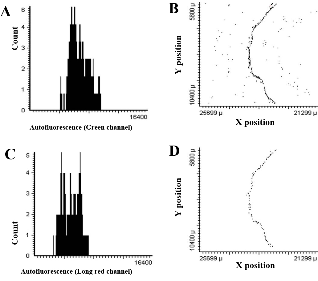
 Figure 9 of
Hjelmeland, Mol Vis 2010; 16:1108-1121.
Figure 9 of
Hjelmeland, Mol Vis 2010; 16:1108-1121.