Figure 8. Double immunolabeling in the C57BL/6J mouse. Detection of retinal pigment epithelium cells double immunolabeled for Mn-superoxide
dismutase and retinal pigment epithelium-specific protein 65 kDa (RPE65) in sections from the posterior pole of a C57BL/6J
mouse by laser scanning cytometry. Panel A shows RPE65 immunolabeling in the green channel (6 μg/ml mouse antibovine RPE65 monoclonal antibody; 7 μg/ml Alexa Fluor
488-conjugated donkey antimouse IgG). Panel B shows the same section viewed in the long-red channel. Panel C shows data from a MnSOD+/RPE65+ double-immunolabeled section in the green channel. The red color in region 2 represents RPE cells that are RPE65+MnSOD+, while green represents RPE cells that are RPE65+MnSOD-. Panel D shows the same section in the long-red channel (3 μg/ml goat anti-SOD2 antibody; 40 μg/ml Alexa Fluor 647-conjugated donkey
antigoat IgG secondary antibody). Abbreviations: RPE65+ represents cells expressing RPE65 protein, MnSOD+ represents cells expressing Mn-superoxide dismutase, MnSOD+/RPE65+ represents cells expressing both Mn-superoxide dismutase and RPE65 protein, C57BL/6J represents C57BL/6J strain of mice.
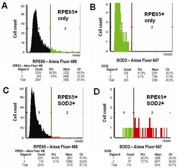
 Figure 8 of
Hjelmeland, Mol Vis 2010; 16:1108-1121.
Figure 8 of
Hjelmeland, Mol Vis 2010; 16:1108-1121.