Figure 7. Double immunolabeling for retinal pigment epithelium-specific protein 65 kDa (RPE65) and Mn-superoxide dismutase (MnSOD).
Detection of retinal pigment epithelium cells double immunolabeled for Mn-superoxide dismutase and RPE65 in sections from
the posterior pole of a 6- and 24-month-old BALB/c mouse by laser scanning cytometry. Panel A shows retinal pigment epithelium RPE65+ cells for the 6-month-old animal. Panel B shows Mn-superoxide dismutase MnSOD+/RPE65+ double-immunopositive cells for a 6-month-old animal. Panel C shows RPE65+ cells from a 24-month-old animal. The red color in region 2 represents RPE cells that are RPE65+MnSOD+, while green represents RPE cells that are RPE65+MnSOD-. Panel D shows MnSOD+/RPE65+ double-immunopositive cells. In each panel, fluorescence intensities are divided between two regions (1 and 2) defined by
a gate at the indicated fluorescence intensity. Each panel shows statistics, including cell counts in each region (Count),
total cell count (Total), percentage of cells in each region (Pct.), Mean (MFI), and coefficient of variation (CV). (Primary
antibodies: 6 μg/ml mouse monoclonal antibovine RPE65 and 3 μg/ml goat anti-SOD2; Secondary antibodies: 4 μg/ml Alexa Fluor
488-conjugated donkey antimouse IgG and 40 μg/ml Alexa Fluor 647-conjugated donkey antigoat IgG). Abbreviations: RPE65+ represents cells expressing RPE65 protein, MnSOD+/RPE65+ represents cells expressing both Mn-superoxide dismutase and RPE65 protein, BALB/c represents BALB/c strain of mice.
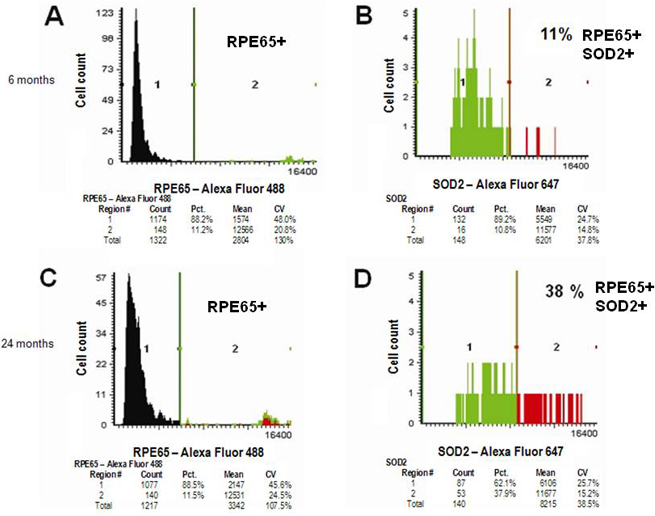
 Figure 7 of
Hjelmeland, Mol Vis 2010; 16:1108-1121.
Figure 7 of
Hjelmeland, Mol Vis 2010; 16:1108-1121.