Figure 6. RPE65 immunolabeling of retinal pigment epithelium (RPE) cells. Identification of retinal pigment epithelium cells by immunolabeling
with an anti-RPE65 monoclonal antibody using laser scanning cytometry. Panel A shows a laser scanning cytometer analysis of a control section (6 μg/ml mouse monoclonal IgG1; 4 μg/ml Alexa Fluor 488-conjugated donkey antimouse IgG secondary antibody). Panel B shows an anti-RPE65 monoclonal antibody immunolabeled section (6 μg/ml mouse antibovine RPE65 monoclonal primary antibody;
4 μg/ml Alexa Fluor 488-conjugated donkey antimouse IgG secondary antibody). The true RPE65+ cells are labeled green. Panel C shows an X–Y plot of all cells, including RPE65+ cells, while panel D shows only RPE65+ cells (arrows). Abbreviations: anti-RPE65 represents antibody against RPE65 protein, IgG represents Immunoglobulin gamma,
RPE65+ represents retinal pigment epithelial cells expressing RPE65 protein.
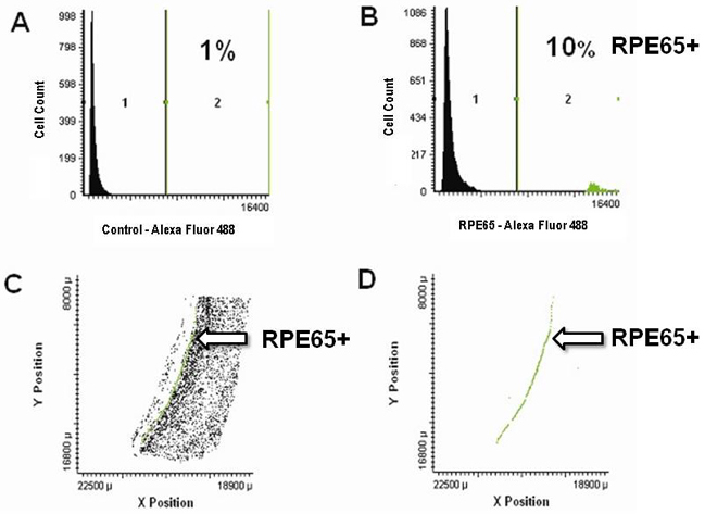
 Figure 6 of
Hjelmeland, Mol Vis 2010; 16:1108-1121.
Figure 6 of
Hjelmeland, Mol Vis 2010; 16:1108-1121.