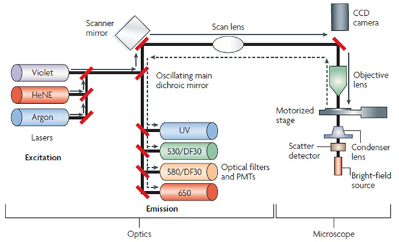
Figure 3. Laser scanning cytometer schematic diagram. The laser scanning cytometer consists of an optics unit that generates the laser
scanning beam, an upright epifluorescence microscope with a motorized stage to allow generation of sample scan images, and
a computer to acquire and analyze scan data using WinCyte software. Fluorescence is excited by laser sources consisting of
an argon laser (blue light; 488 nm), a helium–neon laser (HeNe; red light; 633 nm), and a violet diode laser (405 nm) [
35].
 Figure 3 of
Hjelmeland, Mol Vis 2010; 16:1108-1121.
Figure 3 of
Hjelmeland, Mol Vis 2010; 16:1108-1121.